Immune Checkpoint Inhibitors for Non-Small-Cell Lung Cancer with Brain Metastasis : The Role of Gamma Knife Radiosurgery
Article information

Abstract
Objective
Immune checkpoint inhibitors (ICIs) are approved for treating non-small-cell lung cancer (NSCLC); however, the safety and efficacy of combined ICI and Gamma Knife radiosurgery (GKS) treatment remain undefined. In this study, we retrospectively analyzed patients treated with ICIs with or without GKS at our institute to manage patients with brain metastases from NSCLC.
Methods
We retrospectively reviewed medical records of patients with brain metastases from NSCLC treated with ICIs between January 2015 and December 2017. Of 134 patients, 77 were assessable for brain responses and categorized into three groups as follows : group A, ICI alone (n=26); group B, ICI with concurrent GKS within 14 days (n=24); and group C, ICI with non-concurrent GKS (n=27).
Results
The median follow-up duration after brain metastasis diagnosis was 19.1 months (range, 1–77). At the last follow-up, 53 patients (68.8%) died, 20 were alive, and four were lost to follow-up. The estimated median overall survival (OS) of all patients from the date of brain metastasis diagnosis was 20.0 months (95% confidence interval, 12.5–27.7) (10.0, 22.5, and 42.1 months in groups A, B, and C, respectively). The OS was shorter in group A than in group C (p=0.001). The intracranial disease progression-free survival (p=0.569), local progression-free survival (p=0.457), and complication rates did not significantly differ among the groups. Twelve patients showed leptomeningeal seeding (LMS) during follow-up. The 1-year LMS-free rate in treated with ICI alone group (69.1%) was significantly lower than that in treated with GKS before ICI treatment or within 14 days group (93.2%) (p=0.004).
Conclusion
GKS with ICI showed no favorable OS outcome in treating brain metastasis from NSCLC. However, GKS with ICI did not increase the risk of complications. Furthermore, compared with ICI alone, GKS with ICI may be associated with a reduced incidence of LMS. Further understanding of the mechanism, which remains unknown, may help improve the quality of life of patients with brain metastasis.
INTRODUCTION
Brain metastasis, a common complication of non-small-cell lung cancer (NSCLC), has incidence rates of 30–50% and significantly affects the quality of life of patients [18,49]. The prognosis of patients with brain metastases from NSCLC is extremely poor. Without treatment, the median survival is only 1–2 months. Primary approaches to the treatment of brain metastases include whole-brain radiation therapy, surgery, and stereotactic radiosurgery (SRS) such as Gamma Knife radiosurgery (GKS). Currently, biomarker target agents have led to improved progression-free survival (PFS) and overall survival (OS) in patients with advanced NSCLC [29,33]. Biomarker target agents are a new strategy for treating NSCLC. Some studies have suggested that SRS improves OS and local control in patients with brain metastases [10,24,27,34]. However, these targeted therapies have their own limitations [38,50].
Recently, immune checkpoint inhibitors (ICIs) have been introduced for treating NSCLC. Several drugs, such as pembrolizumab and nivolumab, have been approved for treating NSCLC and malignant melanoma. For both these diseases, clinical trials have shown greater improvement in OS with ICIs than with the standard treatment [4,5,17]. However, there are only few reports on the use of ICIs for treating brain metastases from NSCLC. Although the use of SRS has improved the control of brain lesions and has prolonged survival [9], the efficacy of SRS and ICIs in patients with brain metastases from NSCLC has not been established. In this study, we retrospectively analyzed patients treated with ICIs with or without GKS at our institute to manage patients with brain metastases from NSCLC.
MATERIALS AND METHODS
The study protocol was reviewed and approved by the Institutional Review Board of Samsung Medical Center (SMC-2017-10-051) and was conducted according to the recommendations of the Declaration of Helsinki for biomedical research involving human subjects (1975).
We retrospectively reviewed medical records of patients who were prescribed ICIs (nivolumab and pembrolizumab) for treating NSCLC between January 2015 and December 2017 at our institute. In total, 622 patients were prescribed ICIs, of whom 134 were diagnosed with brain metastasis. Among the 134 patients, 57 lacked follow-up imaging until the last checkup or died and thus were excluded. We enrolled the remaining 77 patients who had follow-up imaging to assess the treatment response. The patients were categorized into three groups based on the administered medications and GKS : group A received ICI alone without GKS, group B received ICI with concurrent GKS within 14 days of prescribing ICI, and group C received ICI with non-concurrent GKS after 14 days of prescribing ICI (Fig. 1).
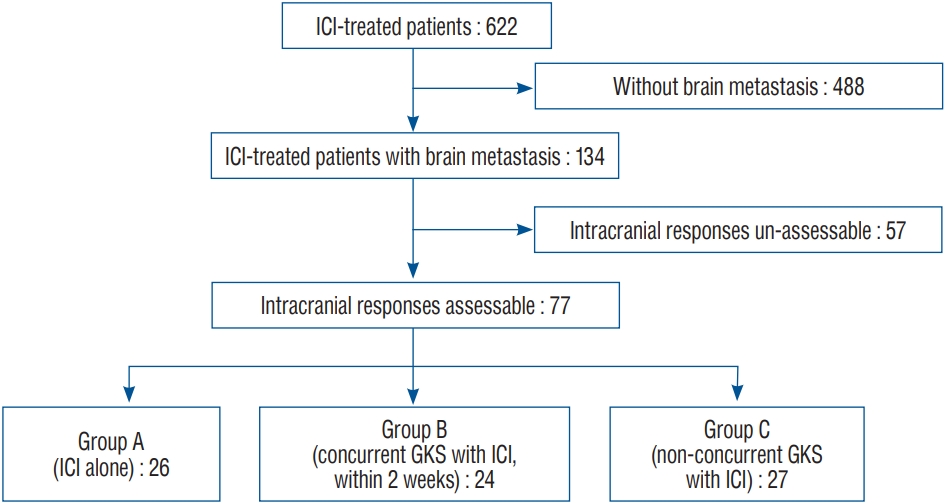
Flow chart of the study. ICI : immune checkpoint inhibitor, GKS : Gamma Knife radiosurgery.
Medication
ICIs (pembrolizumab and nivolumab) were prescribed by neuro-oncologists. The patients received doses of 2 mg/kg pembrolizumab or 3 mg/kg nivolumab every 3 weeks infused intravenously for 30–60 minutes.
Radiosurgical technique
GKS was performed by a neurosurgeon using a Leksell Gamma Knife model Perfexion or ICON (Elekta, Stockholm, Sweden). The median marginal dose of 19 Gy (range, 12–25) at a 50% isodose of the maximum dose was prescribed.
Radiological evaluation
Radiological images obtained using brain magnetic resonance imaging (MRI) were assessed by neuro-radiologists. Local progression of intracranial lesions was defined by the radiological progression of a previously treated metastatic lesion. Intracranial disease progression was defined by the radiological progression of the intracranial condition, including local progression and development of new lesions. Leptomeningeal seeding (LMS) was defined by radiologically identified leptomeningeal progression. To rule out the pseudoprogression by radiation, perfusion images were performed on all MRI.
Statistical analysis
The patients’ demographic and clinical data are summarized using standard descriptive statistics and frequency tabulations. Fisher’s exact test was used to evaluate the differences in categorical variables, and the Kruskal-Wallis test was used to evaluate the differences in continuous variables among the groups. OS was analyzed from the date of diagnosis of brain metastasis to the date of death of the patient. Intracranial disease PFS (I-PFS) was analyzed from the date of initiating treatment with ICI to the date when the intracranial disease progression was revealed using MRI. Local PFS (L-PFS) was analyzed from the date of initiating treatment with ICI to the date when local progression was revealed using MRI. LMSPFS was analyzed from the date of initiating treatment with ICI to the date when LMS was identified. OS, I-PFS, L-PFS, and LMS-PFS were analyzed among the three groups using the log-rank test with Kaplan-Meier plots. p-values <0.05 were considered statistically significant. All analyses were performed in R version 3.4 (https://cran.r-project.org/).
RESULTS
Characteristics of patients and lesions
Seventy-seven patients were enrolled in our study. The median age of these patients was 60 years (range, 42–79) at the time of the diagnosis of brain metastasis; 55 patients were men and 22 were women. The median follow-up duration was 19.1 months (range, 1–77). At the last follow-up, 53 patients (68.8%) died, 20 were alive, and four were lost to follow-up. Adenocarcinomas were identified in 68 patients (88.3%), squamous cell carcinomas in eight (10.4%), and large cell carcinomas in one (1.3%). The mode of onset of brain metastasis was synchronous in 40 patients (51.9%) and metachronous in 37 (48.1%). Nivolumab was prescribed for 31 patients and pembrolizumab for 46 patients. The expression of programmed death-ligand 1 (PD-L1) was assessed in certain patients. The percentage of tumor cells with a level of membrane positivity of >50% was considered positive for PD-L1 protein expression. Thirty-four patients (44.2%) tested positive, 23 (29.8%) tested negative, and 20 did not undergo the evaluation. Ten patients (13.0%) underwent whole-brain radiation therapy before starting ICI treatment. During the follow-up period, 51 patients underwent GKS.
The patients were categorized into three groups based on the administered medications and GKS. Twenty-six patients were included in group A (patients received ICI alone and did not undergo GKS), 24 in group B (ICI with concurrent GKS within 14 days of prescribing ICI), and 27 in group C (ICI with non-concurrent GKS after 14 days of prescribing ICI). The number of brain lesions was counted at the time of diagnosing brain metastasis. The presence of more than 10 lesions and LMS was assigned a value of 10. The median value was 2 (range, 1–10) in all three groups. There were no statistically significant differences in the number of lesions among the three groups (p=0.068) and the patients’ Eastern Cooperative Oncology Group performance status among the three groups (p=1.000). The follow-up duration was shorter in group A than in group B or C (p<0.001). The other baseline characteristics were not statistically different among the three groups (Table 1).
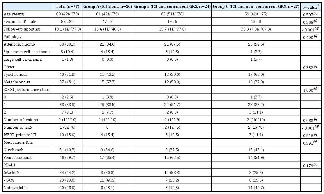
Demographic characteristics of the patients and comparison among the Groups according to differences in the treatment scheme
Survival of patients and disease progression
The estimated median OS of all patients from the date of brain metastasis diagnosis was 20.0 months (95% confidence interval [CI], 12.5–27.7) (estimated median OS were 10.0, 22.5, and 42.1 months in groups A, B, and C, respectively). The OS was significantly shorter in group A than in group C (p=0.001). The estimated median I-PFS from the date of initiating ICI was 4.8 months (95% CI, 2.1–7.5). There were no statistically significant differences in I-PFS among the three groups (p=0.569) (estimated median I-PFS were 3.4, 4.7, and 7.9 months in groups A, B, and C, respectively). The estimated median L-PFS from the date of initiating ICI was 24.0 months (95% CI, 6.9–41.1). There were no statistically significant differences in L-PFS among the three groups (p=0.457) (L-PFS was not reached in group A; the estimated median L-PFS were 26.8 and 11.5 months in groups B and C, respectively) (Fig. 2). During the follow-up period, 53 patients experienced intracranial disease progression. LMS occurred in eight, two, and no patient in groups A, B, and C, respectively. Local treatment failures were observed in one, seven, and two patients in groups A, B, and C, respectively.
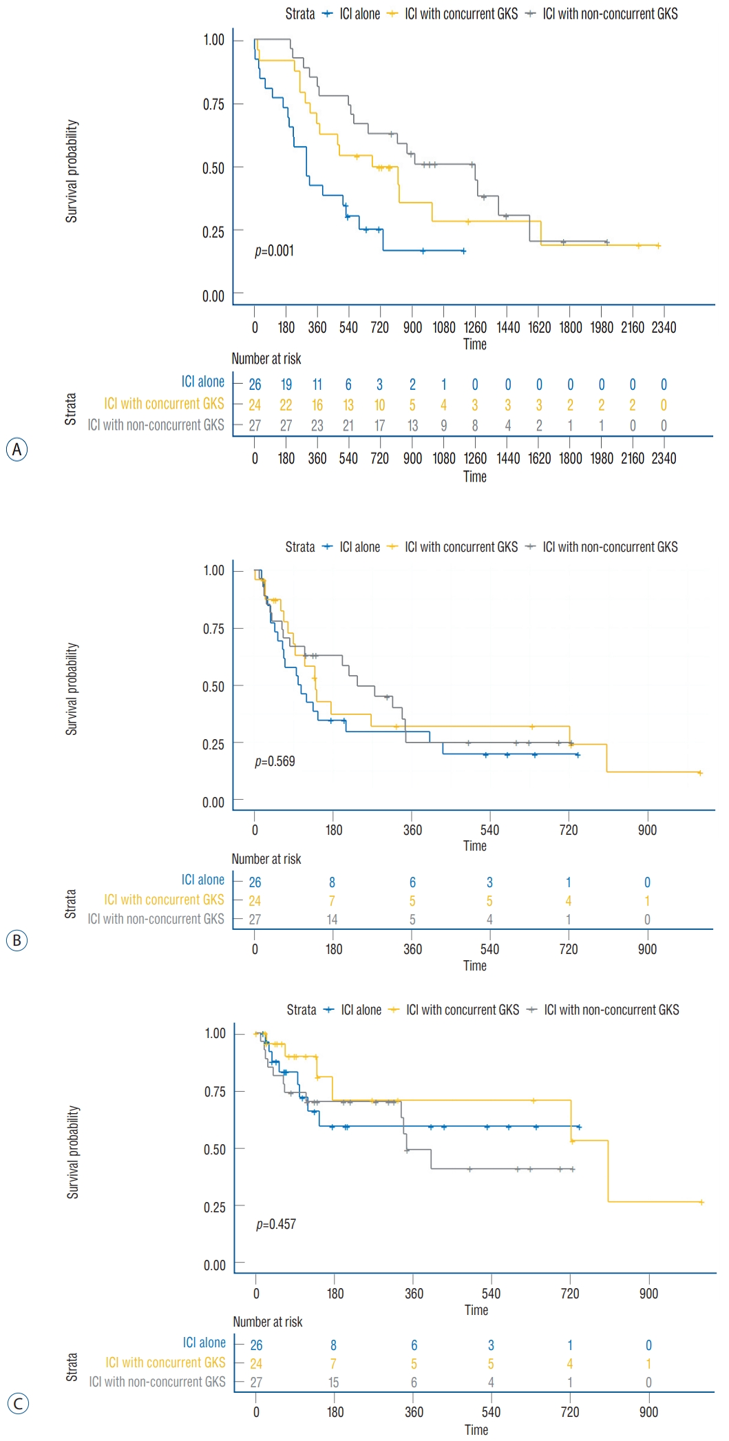
Survival curve according to the treatment group. A : Overall survival. B : Intracranial disease progression-free survival. C : Local progression-free survival according to the treatment group (group A, ICI alone without GKS; group B, ICI with concurrent GKS within 14 days of prescribing ICI; and group C, ICI with non-concurrent GKS after 14 days of prescribing ICI). ICI : immune checkpoint inhibitor, GKS : Gamma Knife radiosurgery.
Effect of ICIs with GKS on LMS
We analyzed the effects of ICI and GKS on the development of LMS. Four patients with LMS diagnosed before starting ICI were excluded, and 73 patients were finally analyzed (24, 23, and 26 patients in groups A, B, and C, respectively). LMS occurred in eight, two, and two patients in groups A, B, and C, respectively. The duration from the initiation of ICI to the development of LMS occurred with a statistically significant difference among the three groups (p=0.015) (the 1-year LMS-free rates were 69.1%, 90.0%, and 95.7% in groups A, B, and C, respectively). The difference was more significant when analyzed from the time of the diagnosis of brain metastasis (p<0.001) (the 1-year LMS-free rates were 74.2%, 95.2%, and 100.0% in groups A, B, and C, respectively).
In group C, only two patients underwent GKS at 2 weeks after completing ICI treatment. We excluded these two patients and analyzed the data again. The results were clearer when the patients were categorized with respect to treatment with ICI alone group and treatment with GKS before ICI treatment or within 14 days. The duration from the initiation of ICI to the occurrence of LMS was statistically significant between the two groups (p=0.004) (the 1-year LMS-free rates were 69.1% and 93.2%, respectively). The duration from the diagnosis of brain metastasis to the occurrence of LMS was also statistically significant among the three groups (p<0.001) (the 1-year LMS-free rates were 74.2% and 97.9% in groups A and B, respectively). The incidence of LMS was lower when ICI was used with GKS (Fig. 3).
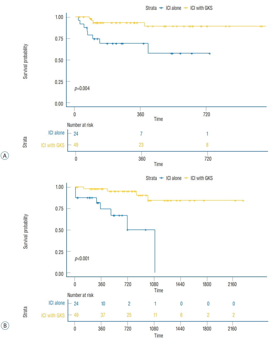
Leptomeningeal progression-free survival curve according to the treatment group duration from the start of ICI treatment (A) and diagnosis of brain metastasis (B) to the development of leptomeningeal seeding (ICI alone without GKS, treatment with GKS before ICI treatment or within 14). ICI : immune checkpoint inhibitor, GKS : Gamma Knife radiosurgery.
Complications
Adverse events were reported according to the National Cancer Institute Common Toxicity Criteria, which were examined during treatment with ICI. There was no adverse event with grade >3. Additionally, there was no case with significant morbidity or mortality related to the treatment (ICI and GKS).
DISCUSSION
ICIs have impressive and long-lasting therapeutic effects on extracranial melanomas and lung cancers. In recent studies, the use of ICI targeting programmed death (PD)-1 and PD-L1 represented a paradigm shift in the management of NSCLC. Some studies have reported significant improvements in OS or quality of life of patients with advanced NSCLC after treatment with ICI compared with those after treatment with other drugs [3-6,11,19,25,45]. The 5-year update of CA209-003 revealed the use of nivolumab in patients with advanced NSCLC resulted in a four-fold improvement in the 5-year OS compared with previous data [19]. Furthermore, in patients treated with pembrolizumab, the 5-year OS was reported to be 23.2% in the treatment-naive group and 15.5% in the pretreated group [11]. The safety of ICI combined with other drugs has also been reported to some extent [16,32]. However, the efficacy of ICI in treating brain tumors needs to be elucidated. The permeability of different drugs through the blood-brain barrier varies among patients and also varies based on the different metastatic lesions in the same patient [36]. ICIs do not directly act on tumors but activate peripheral T cells [41]. Thus, even if the blood-brain barrier is intact, T cells can access the tumor. Furthermore, although normal brain parenchyma or primary brain tumors have immunoregulatory environments with a few infiltrated lymphocytes, metastatic brain lesions have a significant number of tumor-infiltrating lymphocytes [1]. Hence, we assumed that ICIs could be effective in treating metastatic brain tumors. However, there is limited evidence regarding the efficacy of ICIs in treating brain lesions. Preliminary results suggest that ICIs are effective in treating brain metastases in certain patients with NSCLC who were treated with pembrolizumab without radiation therapy [21]. In a recent follow-up study, the response rate of brain lesions was 29.4%, along with a median OS of 8.9 months, with 31% of the patients living for >2 years [22]. Furthermore, the median PFS and OS were 5.5 and 6.5 months, respectively, when patients with brain metastasis from squamous cell lung carcinoma were treated with nivolumab [2]. Another study reported a sudden progression of brain lesions after administering ICIs [28].
The efficacy of radiosurgery with ICI remains unclear. Several reports have revealed that brain metastasis from melanoma and NSCLC has a favorable survival outcome when treated with ICI and SRS [8,30,31,40,43,46-48]; however, some studies have reported no difference in disease control or survival [12,39,42]. In a recent meta-analysis, the application of concurrent radiation along with ICI within 4 weeks showed good results with respect to 12-month OS [35,37]. Our results, however, showed no difference in survival between the concurrent- and non-concurrent-treated groups. The same results were obtained when concurrent was defined as within 2 or 4 weeks. Regarding safety issues, some studies have shown exacerbation of peritumoral edema with the concurrent use of ICI and GKS [14,30], although this was not observed in our study. According to a recently released meta-analysis, a combination of SRS and ICI did not appear to be associated with untoward rates of radionecrosis [35].
Radiation augments antitumor immune responses by releasing tumor antigens and increasing tumor mutational burden following necrosis of tumor cells, thereby causing the release of immune-stimulatory damage-associated molecular patterns. This promotes the recruitment of antigen-presenting cells to the tumor microenvironment [26,51]. Radiation increases PD-L1 expression [13,15]. This suggests that radiation increases the effectiveness of ICI therapy. Chen et al. [8] reported a reduced incidence of new brain metastasis after concurrent SRS, which was explained as a reflection of improved extracranial disease control rather than an abscopal effect. However, our results showed that compared with ICI alone, GKS along with ICI could reduce the incidence of LMS. This could not be explained by extracranial disease control solely. We report here for the first time that SRS delayed the development of LMS in patients with NSCLC treated with an ICI. LMS occurs when malignant cells spread to the cerebrospinal f luid (CSF) space [20,23]. The malignant cells can reach the CSF space via a hematogenous route through the vessels of the arachnoid or choroid plexus [7]. They can also spread from the parenchymal tumor itself while growing or during surgery. LMS develops in 3–5% of patients with advanced NSCLC [44]. LMS may involve a different metastatic process from that of parenchymal metastasis. Our previous study revealed that most patients with brain metastasis from NSCLC experienced LMS in the terminal stage, resulting in a very poor prognosis [9]. This study demonstrated that the early use of ICIs before or with GKS could reduce the incidence of LMS. Unfortunately, we could not completely identify the mechanism of these effects; however, this treatment strategy could aid in improving the quality of life of terminally ill patients with NSCLC.
Because of the retrospective design, our study had some limitations. In total, 42.5% (57/134) of the patients were excluded owing to a lack of clinical information necessary to assess intracranial response. Because they died before we could obtain additional brain imaging, we could not predict the response in these patients. In addition, in the treatment scheme, the prescription of ICI was heterogeneous in a small number of patients with a short follow-up duration. This could make generalization of our results difficult.
Recent studies on the effectiveness of ICI have been published; however, we could not determine a clear factor that could identify patients for whom ICI treatment could be effective. In addition, although many complications caused by ICIs have been reported, there were no specific complications in the current study.
CONCLUSION
The effects of ICIs on the central nervous system, previously considered an immuno-privileged area, have been reported. In this study, ICIs in combination with GKS showed no favorable OS outcome in treating patients with brain metastasis from NSCLC. However, GKS with ICI did not increase the risk of complications. Furthermore, GKS with ICI might be associated with a reduced incidence of LMS; however the exact mechanism for this remains unknown. LMS has a significant impact on the quality of life of end-of-life patients with brain metastasis. It is expected that further understanding of the mechanism may help improve the quality of life of patients with brain metastasis. Further studies are required to identify clinical and molecular predictors to improve the outcomes of patients with brain metastases from NSCLC and to provide clinically meaningful treatment options.
Notes
CONFLICTS OF INTEREST
No potential conflict of interest relevant to this article was reported.
INFORMED CONSENT
This type of study does not require informed consent.
AUTHOR CONTRIBUTIONS
Conceptualization : JIL
Data curation : MHL, KRC, JWC, DSK, HJS, DHN, HAJ, JMS, SHL, JSA, MJA, KP
Formal analysis : MHL
Methodology : MHL
Project administration : JIL
Visualization : MHL
Writing - original draft : JIL
Writing - review & editing : JIL, MHL, KRC, JWC, DSK, HJS, DHN, HAJ, JMS, SHL, JSA, MJA, KP