Congenital Osseus Bridging of Lumbar Transverse Processes
Article information
Abstract
Osseous bridging between lumbar transverse processes is an uncommon condition that may cause low back pain. In most cases, its etiology is alleged to be trauma to the back and only rarely has a congenital origin been indicated. Furthermore, most reported cases involved adults, the majority of whom were middle-aged. Here, the authors describe the case of the youngest girl reported to date with congenital transverse process bridging. As far as the authors' knowledge, there has been no report of congenital bridging of transverse processes in children or adolescents in Korea.
INTRODUCTION
Osseous bridging of adjacent lumbar transverse processes has been reported as a rare complication following trauma to the back in adults2,6). After trauma, heterotopic ossification develops in surrounding soft tissues, and can lead to bridging or pseudarthrosis of transverse processes4,5). In the literature, congenital bridging has been reported much less frequently than traumatic bridging and usually these reports concern adults. Here, we present the simple radiographic and computed tomographic findings of a youngest girl with congenital transverse process bridging. The clinical features of this rare anomaly are discussed with a review of the literature.
CASE REPORT
A 13-year-old girl was referred to our hospital due to low back pain (LBP) and stiffness in the left lower back region. The pain had gradually increased in severity over 1 year, and at presentation she was unable to sit comfortably. She reported that the pain was aggravated by extension of the lower back and prolonged by hours of standing. Her detailed medical history was unremarkable, and there was no history of back injury. A deep, firm fullness was palpable in the paravertebral muscles in the left lumbar area, and was tender to percussion. Lumbar motion was limited in all directions, but the findings of a neurological examination, including the straight leg-raising test (SLRT) were within normal limits. An anteroposterior simple radiographs of the lumbar spine demonstrated the presence of osseous bridging between the left transverse processes of the third and fourth lumbar vertebrae with minimal scoliosis at the bridging site and simultaneous disc space narrowing at the L3-L4 level (Fig. 1). Computed tomography revealed articulation between left transverse processes of the third and fourth lumbar vertebrae (Fig. 2). The left lateral contour of the vertebral body and the bridging transverse processes resembled the shape of the letter 'O', and the components of bone bridge had smooth, regular contours. Because of her young age and parents' wishes, the patient was treated conservatively, that is, by medical and physical therapy. After 6 months of follow up, although her symptoms were still present, they had ameliorated in some degree.
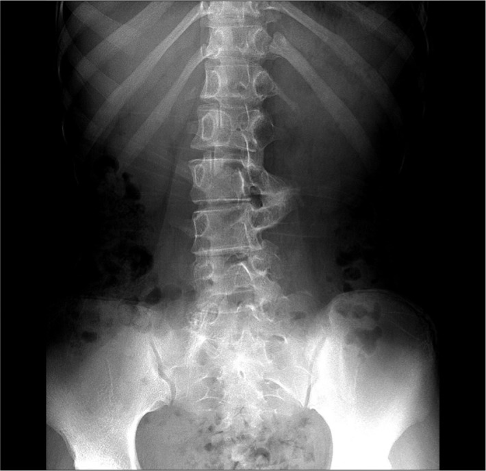
Simple radiograph of the patient. Simple anteroposterior radiograph shows an osseous bridging between left transverse process of L3 and L4 vertebrae and slight scoliosis with disc space narrowing.

Computed tomographic scans of the patient. Coronal and sagittal computed tomographic scans reveal 'O' shaped bridging between transverse processes of L3-L4 vertebrae.
DISCUSSION
Osseous bridging between two transverse processes of the lumbar spine mostly has a traumatic origin in middle aged patients3). Yoslow et al.7), in their review of lumbar osseous bridging in the literature, found that an association exists with back injury and that pseudarthrosis may form in heterotopic bone. In 1991 Billet et al.1) also found that only 15% of their cases had a congenital etiology. In this study, the youngest patient treated was 23 year old, but the origin of the osseous bridge was not obvious. In addition, criteria were suggested for the differentiation of congenital and traumatic osseous bridges. The localizations of hematoma and myositis ossificans play important roles during bony bridge modeling, and the configurations and shapes of traumatic osseous transverse process bridges show acute angles and irregular, asymmetric outlines. On the other hand, osseous bridges of a congenital-developmental etiology are 'O' shaped with symmetrical, contour with convexity of the lumbar spine on the contralateral side and the absence of degenerative changes in the corresponding intervertebral space. Other congenital skeletal anomalies, such as, spina bifida occulta, sacralization of the fifth lumbar vertebra may accompany congenital bone bridges but these findings are not obligatory6). In the present case, the patient had experienced no evident traumatic episode before admission and had a symmetrical smooth regular contour, which was considered indicative of a congenital-developmental etiology. Bone bridging is usually asymptomatic, but low back pain is the most frequent symptom when it becomes symptomatic. Although, the exact mechanism of pain in bony bridge is still not clear, we believe congenital bone union itself, radiculopathy or spinal deformity caused by imbalanced mechanical stress, can contribute to manifestation of symptoms. Our patient showed motion restriction and back pain, but surgical management was not attempted due to improvement of symptoms after medical and physical therapy. Restricted motions of adjacent joints are known to be ameliorated by freeing muscles from the heterotopic mass and by subsequent physical therapy. However, when symptoms are not relieved by conservative treatment, surgical removal of pseudarthrosis should be considered2). The elimination of pain following pseudarthrosis excision is related to local denervation, increased lumbar motion, and the re-establishment of pseudarthrosis in a plane that does not restrict lumbar motion. It would seem appropriate to remove bony bridge when magnetic resonance image or discography fails to show any significant abnormality of the intervertebral disc. We present the imaging findings of this rare anomaly in a young girl with LBP. This is the youngest case reported to date of congenital bridging of transverse processes without back injury.
CONCLUSION
Although rare, this rare anomaly should be borne in mind when evaluating the lumbar spine for the etiology of LBP even in children or adolescents.