Upper Cervical Subluxation and Cervicomedullary Junction Compression in Patients with Rheumatoid Arthritis
Article information
Abstract
Objective
Rheumatoid arthritis (RA) is known to involve the cervical spine up to 86%. It often causes cervical instability like atlantoaxial subluxation (AAS), subaxial subluxation, and vertical subluxation (VS). In order to find the relation between RA and cord compression, we will evaluate the characteristics and risk factors of basilar invagination (BI) and cervicomedullary junction (CMJ) compression.
Methods
From January 2007 to May 2015, 12667 patients administrated to Hanyang University Medical Center. Four thousand three hundred eighty-six patients took cervical X-ray and 250 patients took cervical computed tomography or magnetic resonance imaging. Radiologic parameters, medication records were obtained from 242 patients. Multivariate logistic regression analysis was performed with correlation of CMJ compression, basin-dental interval (BDI), basin-posterior axial line interval (BAI), pannus formation, BI, and AAS.
Results
In the point of CMJ compression, atlantodental interval (ADI), posterior-atlantodental interval, BAI, AAS, and BI are relatively highly correlated. Patients with BI have 82 times strong possibility of radiologic confirmed CMJ compression, while AAS has 6-fold and pannus formation has the 3-fold possibility. Compared to the low incidence of BI, AAS and pannus formation have more proportion in CMJ compression. Furthermore, wrist joint erosion was correlated with VS and AAS.
Conclusion
BI has a very strong possibility of CMJ compression, while AAS and pannus formation have a high proportion in CMJ compression. Hence bilateral wrist joint erosion can be used as an indicator for the timing of screening test for cervical involvement. We suggest the early recommendation of cervical spine examination for the diagnosis of cervical involvement in order to prevent morbidity and mortality.
INTRODUCTION
Rheumatoid arthritis (RA) is a chronic inflammatory polyarthritis disease which mainly involves inflammation of the synovium and enthesis. The incidence of RA has been reported to range from 0.5% to 1% of the adult population [1], which is over 2 million patients with RA in the United States [10,29]. Cervical spine involvement has been reported to be up to 86% of all patients with RA [5,22,39]. RA often causes instability of the cervical spine, which can be categorized into atlantoaxial subluxation (AAS) [13,16,23-26,31,40,44], subaxial subluxation (SAS) [45] and vertical subluxation (VS) [17,32,33]. VS is further classified into cranial settling, vertical migration, atlantoaxial impaction, and basilar invagination (BI). Even though definitions of these terminologies are slightly different, BI is defined as the tip of the odontoid process projects above the foramen magnum, in general.
Due to the low incidence of RA, most studies of cervical involvement in RA have used plain radiography. But a diagnosis of BI by plain radiographs indicates poor sensitivity and a small negative predictive value [36]. In addition, unrecognized cord compression is known to be one of the leading causes of sudden death in patients with RA [28]. In order to find the relation, we will evaluate the characteristics of risk factors of BI and cervicomedullary junction (CMJ) compression in patients with RA statistically using cervical computed tomography (CT) and magnetic resonance imaging (MRI).
MATERIALS AND METHODS
This retrospective study was approved by the Institutional Review Board (IRB) of Hanyang University Medical Center. Due to the retrospective design of the study, consent was neither required by the IRB nor by the study team.
Patient population
Among 12667 individual patients who visited/treated at our institute from January 2007 to May 2015, cervical plain radiographs were obtained from 4386 patients. Plain radiographs were reviewed by radiologists and categorized into three groups as Normal, AAS, and VS. We classified them by the availability of getting cervical CT or cervical MRI data. Finally, 250 patients were investigated in this study.
Medical histories of all patients were undergone full review and radiologic morphologic parameter calculations were performed. Since RA is a progressive autoimmune disease, we considered the most recent CT or MRI as the most disease-progressed image. If a patient exhibited any type of atlantoaxial fixation before the operation, either CT or MRI scans were evaluated. As a result, the preoperative CT or MRI scans of five patients were missing from the data and three patients showed odontoid process fracture with dislocation. These eight patients were excluded from our investigation, which led us to study 242 patients (Fig. 1).
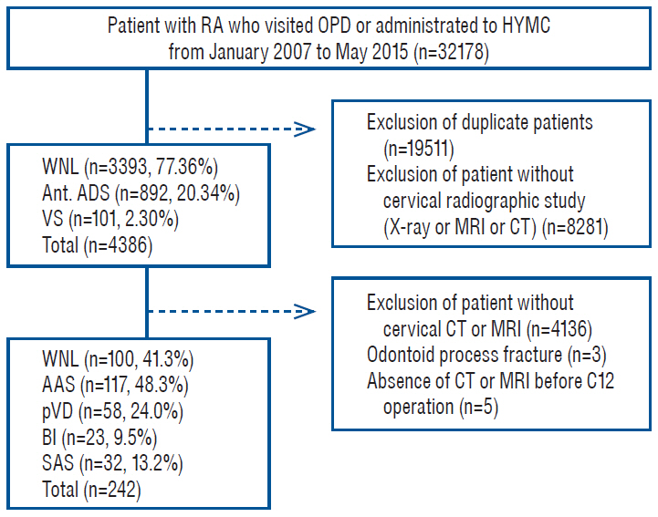
Flow chart of the process for selecting eligible patients for present study. RA : rheumatoid arthritis, OPD : out patient department, HYMC : Hanyang University Medical Center, WNL : with in normal limit, Ant. : anterior, ADS : atlantodental subluxation, VS : vertical subluxation, MRI : magnetic resonance imaging, CT : computed tomography, AAS : atlantoaxial subluxation, pVD : pathologic vertical dislocation, BI : basilar invagination, SAS : subaxial subluxation.
Analysis of cervical CT and MRI
Measurement of radiologic morphologic parameters
Radiologic morphologic parameters were obtained from midsagittal images of cervical CT or MRI scans taken in the neutral position. Atlantodental interval (ADI), posterior-atlantodental interval (PADI), power ratio, the distance between McRae’s line and the tip of odontoid process of the axis (McR), basin-dental interval (BDI), and basion-posterior axial line interval (BAI) were obtained for the coming analysis. A negative value was given if the tip of the odontoid process was below McRae’s line, and positive values were given if the tip of the odontoid process was over the McRae’s line. All the geometrical lengths were measured by J.W.C. and M.H.H., and the mean value was calculated. As the geometrical lengths are quantitive variables (ADI, PADI, power’s ratio, McR, BDI, and BAI), we considered the values agreed if the data differs less than 10%. The interobserver agreement the radiographic parameters varies from 0.76 to 0.87, which are substantial agreement to excellent agreement.
Presence of bony erosion of odontoid process, pannus formation, and SAS was investigated, but subluxation related to trauma history was excluded. CMJ compression was determined from both cervical CT and MRI scans. Impressions of CMJ contours or signal changes and/or syrinx formation in cervical MRI scans were regarded as definite CMJ compression. We also included patients with a simple attachment of the odontoid process tip in the CMJ compression group, since this may potentially lead to compression during cervical movement.
Criteria of atlantoaxial subluxation, pathologic vertical dislocation, and basilar invagination
Since cervical CT and MRI scans were taken in the neutral position, we determined ADI from lateral and flexion/extension views of cervical X-rays. We defined pathological AAS when ADI obtained from cervical radiographic images is greater than or equal to 3 mm. Various methods for measuring VS have been introduced [17,19,27,32-34]; however, the diagnostic criteria for VS are less clear than those of AAS. Since BI is marked out when the tip of the odontoid process axis projects over McRae’s line, we consider McRae’s line as the reference line. Cronin et al. [8] have reported that the mean tip of the odontoid process is 4.6 mm for women and 5.3 mm for men with standard deviation (SD) 1.8 mm below Mc Rae’s line from cervical CT scans. But the mean tip is 4.6 mm in both sexes with median 4.8 mm and SD 2.6 mm below McRae’s line from cervical MRI scans [9]. Thus, pathologic vertical dislocation (pVD) is demarcated when the tip of the odontoid process is located within 3 mm below McRae’s line.
Hand X-ray analysis
Since all patients in this study were diagnosed with RA, most of them, in fact 217 patients, had undergone hand X-rays. The extent of wrist joint bony erosion and the presence of finger deformities were examined. Finger deformity is defined by an angle was greater than 10 degrees between the shafts and any interphalangeal joints or metacarpal phalange joints. But finger deformity due to trauma is excluded in this study.
Medical history review
The medical history of each patient with RA was considered. Since dosage use and period were highly variable, we only considered whether the patients used the medication or not. Medications were categorized into steroids (prednisolone, Calcort, Tracinon), disease-modifying antirheumatic drugs (DMARDs : Haloxin, ARAVA, Zopyrin), immunosuppressants (Methotrexate, Bredinin, Cipol, Azathioprine), and biologic drugs (adalimumab, etanercept, abatacept).
The results of a clinical laboratory test were also investigated. In particular, the erythrocyte sedimentation rate (ESR), level of c-reactive protein (CRP), level of anti-cyclic citrullinated peptide (anti-CCP) antibodies, and level of the rheumatoid factor were obtained adjacent to each patient’s cervical CT or MRI scan. Since ESR and CRP are often elevated in the presence of any kind of inflammation, the median is calculated from the data obtained by each patient’s cervical CT or MRI scan during the preceding 1-year period. We call it high titer when the median is greater than three-fold above the upper limit of the normal value of each lab test.
Statistical analysis
Firstly, Spearman’s Rho test was applied to identify correlations among VS, AAS, and CMJ compression with other variables. The p-values and correlation coefficients (CCs) were calculated for each test. We take statistical significance when p<0.05 and CC ≥0.40. Secondly, univariate logistic regression was used in order to analyze relations among CMJ compression, BI, AAS, and SAS with other variables. Thirdly, multivariable logistic regression was applied to find variables having statistically significant relations. All statistical analysis were performed using commercial software (SPSS Statistics, version 21; IBM, Armonk, NY, USA). Of course, missing data were excluded from each analysis.
RESULTS
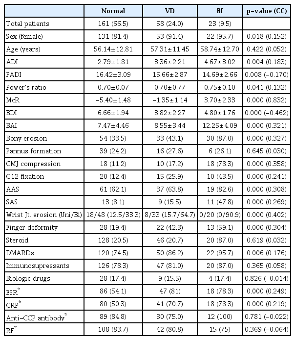
Multiple variables were significantly correlated with VS (pVD and BI). However, most of them are weakly correlated, while McR, BDI, and wrist joint erosion are relatively strongly correlated with VS. Since McR is closely related to the bounds of pVD and BI, their correlations are consistent with the expected results. BDI shows negative correlations with pVD and BI. But pVD and BI are significantly correlated with wrist joint erosion. Therefore, we may say that the proportion of bilateral wrist joint erosion is increasing since the vertical dislocation of the atlantoaxial joint as in Table 1.
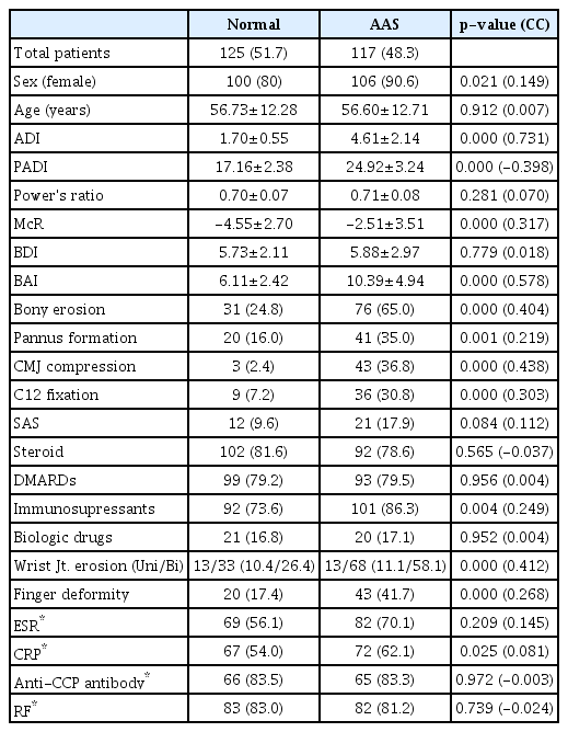
We also analyzed the data in perspective of AAS including ADI, PADI, BAI, bony erosion of the odontoid process, CMJ compression, and wrist joint erosion. In all perspectives, relatively strong significant correlations are derived. ADI, PADI, and BAI are closely related to horizontal dislocation of the atlantoaxial joint, which is coincident with our prediction. Moreover, bony erosion of the odontoid process and bilateral wrist joints have also statistically significant correlation coefficients with AAS. These results indicate that hand X-ray findings can potentially predict AAS. It is well known that AAS amplitude tends to be stabilized or decreasing as VD develops or progresses [6,18,31,38]. McR value is significantly different between normal group and AAS group, however in reveals low CC. This finding suggests that BI can be the result of development and progression of AAS (Table 2).
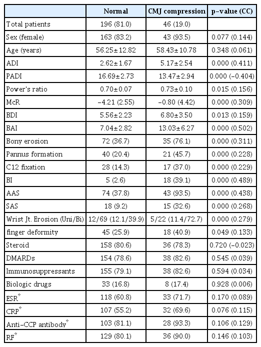
Since ADI, PADI, BAI, AAS, and BI are significantly correlated with CMJ compression, it follows from Table 3 that CMJ compression is related to excess subluxation both horizontally and vertically. In order to determine the most relevant index for diagnosis of CMJ compression, receiver operating characteristics (ROC) curve analysis is applied and the area under the curve (AUC) is calculated, whose results are graphed as in Fig. 2. It follows from Fig. 2 that BAI is the most predictive index for CMJ compression (AUC, 0.869), followed by ADI (AUC, 0.802) and then by McR (AUC, 0.727). Using 12 mm-cutoff of BAI which was described by Harris et al. [14,15] (Harris’s rule of 12’s), the sensitivity is 0.920 (95% confidence interval [CI], 0.893–0.942) and the specificity is 0.692 (95% CI, 0.552–0.805). Moreover, the positive predictive value is 0.939 (95% CI, 0.911–0.961) and the negative predictive value is 0.628 (95% CI, 0.501–0.730).
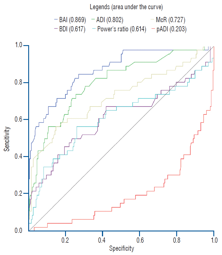
ROC curve analysis was performed in radiologic morphologic parameters. Note that area under the curve of BAI is larger than that of ADI and McR, which indicates higher sensitivity and specificity. BAI : basin-posterior axial line interval, ADI : atlantodental interval, McR : McRae’s line and the tip of odontoid process of the axis, BDI : basindental interval, pADI : posterior-atlantodental interval, ROC : receiver operating characteristics.
Multivariate logistic regression analysis is performed on ADI, PADI, Power ratio, McR, BDI, BAI, bony erosion of the odontoid process, pannus formation, pVD, BI, AAS, SAS, and wrist joint erosion. Regression analysis is carried out by using the backward elimination process with a 95% CI. Table 4 shows that CMJ compression happens approximately 82 times more as in BI when the odontoid process passing over McRae’s line and six times more as in AAS when the horizontal dislocation is observed over 3 mm. Also, pannus formation increases the risk of CMJ compression by 3-fold.
DISCUSSION
AAS is the most common cervical abnormality in patients with RA. It is caused by ligamentous structural destruction of the craniovertebral junction, which results from chronic inf lammation [31]. Secondly, upper spine lesions are commonly involved in the pure synovial component of the occipito-atlanto-axial joint complex, which is the main target involved in RA. The occipital-atlanto-axial joint absorbs most of the rotational movement and is supported only by a ligamentous structure without any interlocking anatomic bony structures. Thus, the loosening of this ligamentous structure can lead to instability and dislocation in any direction [35]. In particular, destruction of the bony compartment of the occipito-atlantoaxial joint complex leads to VS, which is the thirdly most common cervical abnormality in patients with RA after SAS [6,18,35].
It has been reported that 40% to 88% of patients with RA complain of neck pain. Furthermore, cervical spine involvement is reported in 43% to 86% of all RA patients based on radiographic studies, whereas neurologic symptoms occur in 7% to 34% of all patients [3,30]. Under the assumption that the radiographic study of the cervical spine is only performed on patients complaining of significant cervical pain, 34.6% of patients with RA have cervical pain. However, in this investigation, 22.6% of patients with RA have cervical involvement in cervical plain radiographs and 58.7% in cervical CT or MRI scans. Moreover, 2.30% of all cervical plain radiographs show VS, whereas 33.5% of them are seen in cervical CT or MRI scans. According to this result, we may conclude that patients undergone CT or MRI have a higher incidence of cervical spine involvement. This indicates that CT and MRI yield a more accurate resolution in determining the cervical anatomy of VS. However, selection bias toward CT/MRI may also lead to this phenomenon. On the other hand, the proportion of cervical spine involvement can be lower in the whole population, since 50% of all patients are asymptomatic even though they have radiographic instability [7,35].
Numerous methods have been developed to interpret the anatomical status of the occipito-atlanto-axial joint complex [6,12-15,22]. Variables related to VS and AAS are strongly correlated, while others are weakly correlated. Without any of variables related to anatomic relationship with the occipito-atlantoaxial joint complex, Table 2 shows that wrist joint erosion is correlated with VS (p-value <0.001; CC, 0.402) and AAS (p-value <0.001; CC, 0.412). Wrist joints are known to be the most frequently involved joints in RA and are often involved in early disease [12,42,43]. Thus, wrist joints and hands are examined together in the diagnosis of RA, which is a cost-effective approach for the diagnosis. Although the incidence of cervical involvement in RA is relatively low compared to the involvement of hand joints, the morbidity and mortality resulting from cervical involvement can be devastated [28]. Therefore, we recommend the cervical spine evaluation with plain radiograph images at an early stage. A screening test can be also recommended for patients with erosion on their wrist joints.
Patients with CMJ compression having BI may have neurologic symptoms such as intractable pain, motor or sensory neurologic deficits, myelopathy and even sudden death [4,28,37]. The incidence of BI in patients with RA has been reported to range from 5% to 34% [6,20,21,37]. But, in this study, the incidence of BI is less than 2.3% in patients with RA. More surprisingly, 93.5% (43 out of 46 patients) of cases with CMJ compression confirmed radiologically by cervical CT or MRI has AAS, and 39.1% of them has BI. However, multivariate logistic regression indicates that patients with BI are 82 times more likely to have CMJ compression, while AAS and pannus formation had six times and three times more likely, respectively. But AAS and pannus formation are observed relatively often in CMJ compression due to the low incidence of BI.
Since CMJ compression is associated with not only BI but also AAS and pannus formation, we analyze radiologic parameters subsequently. In ROC curve analysis, BAI has the largest AUC (AUC, 0.869). When we use 12 mm-cutoff of BAI, sensitivity and specificity are 0.920 and 0.692, respectively. In the progression of BI, the odontoid process must dislocate posteriorly to pass the foramen magnum. Thus, in BI the BAI must be at least larger than the anterior-posterior diameter of the odontoid process. Since this metric represents both AAS and BI, BAI has more coverage in CMJ compression compared to ADI or McR. Riew et al. [36] have studied that diagnosis of BI in plain radiographs can be problematic since the odontoid process is clearly identified in only 35% of patients. BAI has an advantage as it does not require any identification of the odontoid process. Furthermore, 12 mm-cutoff of BAI yields high sensitivity and a relatively large negative predictive value. It indicates that this parameter can be used to screen for CMJ compression. However, visualization of basion from plain radiographs of RA patients can be often difficult in osteopenia, erosions and conjoined subluxations [11,17,30,32,41]. Thus, additional cervical CT or MRI is required in these patients.
Limitations
There are some limitations to this study. Firstly, patients suspected of RA visit to our institute from all over the nation since the hospital runs an independent specialized hospital for rheumatic disease. This makes it possible to get a large population of patients with RA. This population may lead to having different characteristics compared with those in other studies. Moreover, since relatively early diagnosis and treatment for RA may be possible in the hospital, it may lead to relatively lower incidences of AAS and VS than others. Secondly, this study is limited in its retrospective cross-sectional design even though it is based on a large number of populations. Moreover, it was not able to estimate RA activity during the study. by using DAS/DAS 28 and the patient’s neurologic examination results due to incomplete medical records. Therefore, it may not reveal the pathogenesis mechanisms of either BI or CMJ compression. Thirdly, RA is a chronic autoimmune disorder and some differences may present between the date of diagnosis and the date of RA onset. But it was impossible to narrow down the diagnostic criteria for each individual from the retrospective medical database since we did not consider the change of medical historic data from 1987 American College of Rheumatology (ACR) criteria for RA to 2010 ACR/European League Against Rheumatism (EULAR) classification criteria [2].
CONCLUSION
Cervical spine involvement is one of the most common types of pathogenesis in patients with RA and CMJ compression can lead to devastating outcomes. Our results indicate that BI is associated with a very strong probability of CMJ compression and patients with CMJ compression exhibit high proportions of AAS and pannus formation. Moreover, bilateral wrist joint erosion is a potential indicator for screening for cervical involvement of RA. Thus, we recommend early cervical spine examinations for patients with RA in order to diagnose the extent of cervical involvement. For patients with BAI over 12 mm, cervical CT or MRI should be performed to evaluate the extent of CMJ compression with the goal of preventing morbidity and mortality. A cohort study of cervical spine involvement with detailed neurologic examinations is needed to define the clinical correlations and pathogenesis mechanisms involved in this disease precisely.
Notes
No potential conflict of interest relevant to this article was reported.
INFORMED CONSENT
This type of study does not require informed consent.
AUTHOR CONTRIBUTIONS
Conceptualization : KHB, JC
Data curation : JC
Formal analysis : JC, MHH
Methodology : JC, KHB, MHH
Project administration : KHB, JC
Visualization : JC
Writing - original draft : JC
Writing - review & editing : KHB, HJY, HJC, JIR, MHH

