Can Right-Handed Surgeons Insert Upper Thoracic Pedicle Screws in much Comfortable Position? Right-Handedness Problem on the Left Side
Article information
Abstract
Objective
Thoracic pedicles have special and specific properties. In particular, upper thoracic pedicles are positioned in craniocaudal plane. Therefore, manipulation of thoracic pedicle screws on the left side is difficult for right-handed surgeons. We recommend a new position to insert thoracic pedicle screw that will be much comfortable for spine surgeons.
Methods
We retrospectively reviewed 33 patients who underwent upper thoracic pedicle screw instrumentation. In 15 patients, a total of 110 thoracic pedicle screws were inserted to the upper thoracic spine (T1–6) with classical position (anesthesiologist and monitor were placed near to patient’s head. Surgeons were standing classically near to patient’s body while patients were lying in prone position). In 18 patients, a total of 88 thoracic pedicle screws were inserted to the upper thoracic spine with the new standing position-surgeons stand by the head of the patient and the anesthesia monitor laterally and under patient’s belt level. All the operations performed by the same senior spine surgeons with the help of C-arm. Postoperative computed tomography scans were obtained to assess the screw placement. The screw malposition and pedicle wall violations were divided and evaluated separately. Cortical penetration were measured and graded at either : 1–2 mm penetration, 2–4 mm penetration and >4 mm penetration.
Results
Total 198 screws were inserted with two different standing positions. Of 198 screws 110 were in the classical positioning group and 88 were in the new positioning group. Incorrect screw placement was found in 33 screws (16.6%). The difference between total screw malposition by both standing positions were found to be statistically significant (p=0.011). The difference between total pedicle wall violations by both standing positions were found to be statistically significant (p=0.003).
Conclusion
Right-handedness is a problem during the upper thoracic pedicle screw placement on the left side. Changing the surgeon’s position standing near to patient’s head could provide a much comfortable position to orient the craniocaudal plane of the thoracic pedicles.
INTRODUCTION
Pedicle screws are widely used for treatment of trauma, spinal tumors, deformity, spinal instability and infections. Insertion of pedicle screws in thoracic spine, even in the experienced hands, was noted to have high incidences of pedicle violation [7,10,15,18].
Correct insertion of thoracic pedicle screws is much important in correct position because of the closeness of the spinal cord and relative narrowed spinal canal [10,12]. Moreover, insertion of thoracic pedicle on the left side needs more caution because aorta is placed close to posterolateral side of the vertebral column [8,16]. In particular, upper thoracic pedicles between T1 and T6 are positioned in cranio-caudal plane (Lien). With the classical position, anesthesiologist and monitor were placed near to patient’s head. Surgeons stand classically near to patient’s body while patients are lying in prone position. Manipulation of thoracic pedicle screws on the left side is difficult for right-handed surgeons in classical position [20]. We recommend a new standing position to insert pedicle screws in upper thoracic segments. With the new standing position, surgeons stand by the head of the patient and the anesthesia monitor is placed lateral side, under patient’s belt level (Fig. 1).
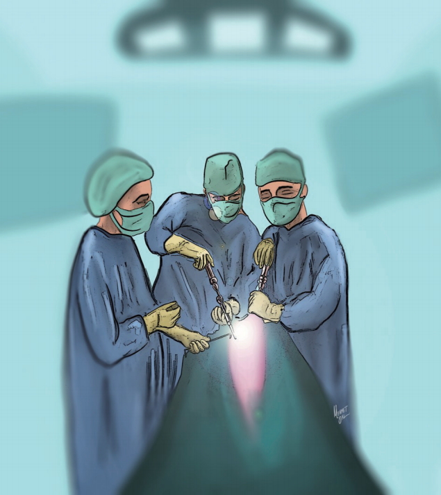
The new standing position: Surgeons stand by the head of the patient and the anesthesia monitor placed laterally, under patient’s belt level.
In this study, we retrospectively evaluated the two different positions for right-handed surgeons during inserting pedicle screws into the upper thoracic spine. Our hypothesis is that the new position we recommended may maintain more comfortable position for the right-handed spine surgeons on the left side.
MATERIALS AND METHODS
Patients who underwent upper thoracic spine pedicle screw placement between 2010 and 2016 were retrospectively evaluated. Patients’ demographics were detailed in Table 1. Patients were divided into two groups named classical positioning group and new positioning group. Both groups divided into two subgroups named screw malposition group and pedicle wall violation group. Finally, pedicle wall violation group divided into four subgroups named medial, lateral, superior and inferior. Postoperative computed tomography (CT) scans of the thoracic spine were obtained for all patients. Pedicle screws were marked as ‘‘correct’’ if they were fully contained within the pedicle walls. Besides, pedicle wall violations were measured and graded according to Gertzbein classification at either : 1–2 mm penetration, 2–4 mm penetration, or greater than 4 mm penetration [5]. Right or left sides of the cortical penetrations were marked. The screw malposition and pedicle wall violations were divided and evaluated separately.
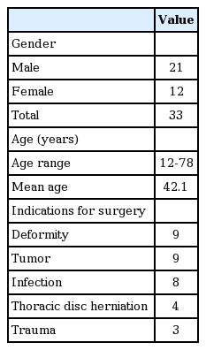
Patient demographics and indications for surgery
Standing positions
Patients were operated under general anesthesia by two right-handed spine surgeons. In the first group, anesthesiologist and monitor were placed near to patient’s head. Surgeons stood classically lateral sides of the patient’s body while patients were lying in prone position. In the second group, surgeons stood by the head of the patient and the anesthesia monitor was placed on the lateral side, under patient’s belt level (Fig. 1). For the both groups, the thoracic spinal level was confirmed under per-operative C-arm fluoroscopy with caution. The sagittal trajectory of the probe is based upon the external anatomy of the posterior thoracic spine, the lamina and spinous processes. Cranio-caudal angulation is based upon per-operative C-arm flouroscopy. Free-hand technique [10] was used when inserting the screws. 4.0–4.5 mm diameter pedicle screws were placed between T1–6 for spinal stabilization.
Statistical evaluation
Statistical analyses were conducted using SPSS (IBM Corp., Armonk, NY, USA; IBM SPSS Statistics for Macintosh version 23.0, IBM Corp.). Chi-square test was utilized to check the proportion of significant medial, lateral, superior and inferior pedicle wall violations by both the standing positions for right and left sides. Results are expressed as the mean (range), with a p value of 0.05 considered as being statistically significant.
RESULTS
We retrospectively reviewed 33 patients (21 males and 12 females) who underwent upper thoracic pedicle screw instrumentation due to various pathologies. Mean age was 42.1 years in range between 12 and 78 years. In 15 patients, a total of 110 thoracic pedicle screws were inserted to the upper thoracic spine (T1–6) with classical position. In 18 patients, a total of 88 screws were inserted to the upper thoracic spine with the new position (Fig. 1). Indications for thoracic spine surgery were spinal deformity (n=9, 27%), spinal trauma (n=9, 27%), metastatic or primary tumors (n=8, 25%), thoracic disc herniation (n=4, 12%) and spinal infections (n=3, 9%).
Total 198 screws were inserted to upper thoracic vertebras of the patients. Of 198 screws 110 were in the classical positioning group and 88 were in the new positioning group.
Incorrect screw containments in medial, lateral, superior and inferior directions were assessed by CT scans and results were shown in Table 2. Total screw malposition rate was 16.6% (n=33) in both groups. Eighty-five screws (77.3%) were completely within the pedicle in the classical positioning group (Table 2). Twenty-five screws (22.7%) penetrated the pedicle walls according to Gertzbein classification. Six incorrect screws were on the right side, whereas 19 were on the left side. Seventeen screws penetrated in one direction and eight screw penetrated in two directions. In the axial CT images, total 33 pedicle wall violations were observed. Three of the pedicle wall violations were medially, 11 of the pedicle wall violations were laterally, 16 of the pedicle wall violations were superiorly, and three of the pedicle wall violations were inferiorly. Twenty-five of the pedicle wall violations were on the left side and 8 were on the right side (Table 2).
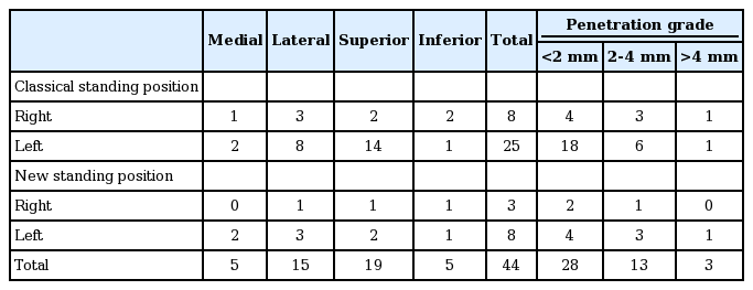
Sides, directions numbers and degrees of the pedicle wall violations with the classical standing position and new standing position
Malposition rate of the screws positioned via the new standing position was 9% (n=8). Three incorrect screws were on the right side, whereas five were on the left side (Table 1). Two screws penetrated in one direction and three screws penetrated in two directions. In the axial CT images, two pedicle wall violations were medially, four pedicle wall violations were laterally, three pedicle wall violations were superiorly, and two pedicle wall violations were inferiorly. Right and left side total pedicle wall violations were three and eight, respectively (Table 2).
The differences between the medial, lateral, and inferior pedicle walls violations by both standing positions were found to be statistically insignificant for the left side. The difference between the superior pedicle wall violations by both standing position was found to be statistically significant the for left side (p=0.004) (Table 3).

p-values by chi-square test for violation in both standing positions by sides and directions
The differences between the medial, lateral, superior, and inferior pedicle walls violations in both two groups were found to be statistically insignificant for the right side. The difference between total pedicle wall violations by both standing positions were found to be statistically signif icant (p=0.003) (Table 4). Likewise, the difference between total screw malposition by both standing positions were found to be statistically significant (p=0.011). The difference between the left side screw malpositions in the new positioning group (n=5) and total right side screw malpositions (n=9) in both groups were found to be statistically insignificant (p=0.827).
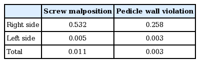
p-values by chi-square test for screw malposition and pedicle wall penetration in both standing positions by sides
Surgeon one and surgeon two inserted 18 of 102 screws incorrectly and 15 of 96 screws incorrectly, respectively. The difference between incorrect screw placements of both surgeons was statistically insignificant (p=0.703).
DISCUSSION
Inserting pedicle screws into upper thoracic spine needs talent and experience. Additionally, the positioning and insertion technique are very important for this challenging intervention. Changing the standing position of surgeons may decrease the malposition rate of right-handed spine surgeons on the left side when inserting pedicle screws into upper thoracic spine.
Thoracic pedicle screws biomechanically maintain stronger stabilization when compared to other fusion techniques. The pullout strength of the pedicle screws is much stronger and the fusion rates are higher due to larger bone-implant area [7,11,14]. Per-operative fluoroscopy has been the most commonly used method to guide the insertion of pedicle screws into thoracic spine. Nevertheless, up to 40% rate of pedicle wall violation has been noted in the literature [19]. The free-hand thoracic pedicle screw technique has been described by Kim and Lenke [10] Pedicle screw insertion based on anatomical landmarks and the tactile feel of probing the pedicles. In this study, the free-hand technique was used to insert pedicle screws into thoracic spine. Suk et al. [17] described biplaner thoracic pedicle technique. Four thousand six hundred four pedicle screws were placed with their technique and 1.5% of the pedicles were violated medially. Of the 279 pedicles screws, only two screws penetrated spinal canal (medial wall) in Belmont series [1]. In our patients, five screws of 198 screws were placed medially (2.5%).
The rate of complications due to incorrect pedicle insertion is between 0% and 0.9% in the literature [1,4,6]. Medial pedicle wall violation can cause cord injury, neurological deficits and CSF leakage due to dural injury. Lateral wall violation can cause vascular injury and, pleural irritation and visceral injury [3]. Superior and inferior walls violation can cause dorsal pain, musculoskeletal pain and rib pain due to thoracic nerve root radiculopathy and CSF leakage [1,3,4]. Moreover, pedicle wall violation causes reduction of pull-out strength of the pedicle screws due to reduced bone-implant interface [2]. In our patients, 33 screws of the 198 thoracic screws were inserted incorrectly (16.6%). However, result of this study correlated with previous studies. Our patients did not have any neurological and vascular complications due to incorrect pedicle screw insertion.
In this study, 25 of the pedicle wall violations were on the left side, eight were on the right side in classical positioning group. Violations of the screws were more frequent on the left side, and the difference was statistically significant (p=0.001). In the new positioning group, eight of the pedicle wall violations were on the left side, three were on the right side. The difference between two sides was statistically insignificant (p=0.170). The difference between total pedicle wall violations by both standing positions were found to be statistically significant (p=0.003). Likewise, the difference between total screw malposition by both standing positions were found to be statistically significant (p=0.011). The difference between the left side screw malpositions in the new positioning group (n=5) and total right side screw malpositions (n=9) in both groups were found to be statistically insignificant (p=0.827). This result supports that new standing position is successful to eliminate the difference between both right and left sides.
Thoracic pedicles have special and specific properties. The pedicles of thoracic vertebra are placed in craniocaudal plane [13]. In their large series, Kim and colleagues recommended that the direction of the pedicle screw should be more caudal and lateral in the upper thoracic region [9]. With the classical standing position, it is difficult to direct the screws to medial and superior due to limitation of the patient’s body on the operating table for the right-handed surgeon on the left side. The difference between the superior pedicle wall violations by both standing positions was found to be statistically significant for the left side (p=0.004). Otherwise, the difference between medial wall violations by both standing positions was found to be statistically insignificant the for left side (p=0.858). This can be explained by the fact that medial wall of thoracic pedicle is stronger than the lateral wall. The screws oriented medially usually don’t penetrate medial wall because of the medial wall’ s plasticity and thickening [10]. In particular, the medial pedicle wall tents to keep in the pedicle due to its strong structure in free-hand technique. Moreover, the desire of the surgeon is usually to avoid potential injury to the spinal cord, thus the surgeons force to insert screw laterally uncomfortably.
In conclusion, the insertion of thoracic pedicle on the right side is easily for right-handed surgeons but insertion of the thoracic pedicle screw on the left side is difficult for righthanded surgeons because the pedicles of thoracic vertebra are placed in cranio-caudal plane. Instead of classical position, placing the anesthesia monitor lateral side of the patient in prone position (under belt level) and maintaining position of the surgeons close to the patient’s head during inserting upper thoracic spine will provide a comfortable position for the right-handed surgeons and decrease the screw malpositioning rate especially on the left side.
This study had some limitations. The number of patients was low. A more cases study should be performed. Secondly, this is a retrospective study in a single center; a multicenter prospective randomized controlled study between classical standing position and new standing position should be performed.
CONCLUSION
Right-handedness is an important problem during the upper thoracic pedicle screw placement on the left side due to specific properties of the pedicles. Changing the surgeon’s position, standing near to patient’s head will provide a much comfortable position to orient during inserting pedicle screw in the upper thoracic spine.
Notes
No potential conflict of interest relevant to this article was reported.
INFORMED CONSENT
This type of study does not require informed consent.