Prevalence of Disc Degeneration in Asymptomatic Korean Subjects. Part 1 : Lumbar Spine
Article information
Abstract
Objective
Asymptomatic patients show high degeneration prevalence at lumbar disc in previous literatures. Unfortunately, there are few Korean data, so the authors attempted to analyze the prevalence of disc degeneration in highly selective asymptomatic Korean subjects using MRI.
Methods
We performed 3 T MRI sagittal scans from T12 to S1 on 102 asymptomatic subjects (50 men and 52 women) who visited our hospital between the ages of 14 and 82 years (mean age 46.3 years). All images were read independently by three observers (two neurosurgeons and one neuroradiologist) who were not given any information about the subjects. We classified grading for lumbar disc herniation (HN), annular fissure (AF), and nucleus degeneration (ND), using disc degeneration classification.
Results
The prevalence of HN, AF, and ND were 81.4%, 76.1%, and 75.8% respectively. Almost all levels showed an age-related proportional tendency with some exceptions.
Conclusion
In asymptomatic Korean subjects, the abnormal findings showed high prevalence of AF, ND, and extrusion. Especially in young ages, the authors found that bulging, protrusion, and AF showed high prevalence at L4/5 and L5/S1. And ND showed high prevalence at L5/S1. So, all lumbar disc degenerations are not pathologic, especially in children and adolescents.
INTRODUCTION
According to literature, the prevalence of lumbar pain at one time point is approximately 30-40% in Germany. The one year and lifetime prevalence are approximately 60-70% and more than 80%, respectively30). In industrialized Korea, low back pain is second only to headache as a cause of pain11). Koreans have traditionally used the Ondol system (traditional floor heating system), so a lifestyle of sitting on the floor such as cross-legged sitting has been the norm with chair sitting only gaining popularity since 1980s. Due to the difference in lifestyle between Koreans and Westerners, there has been an opinion that the prevalence of disc degeneration in the normal population between the two would differ.
Asymptomatic patients show high degeneration prevalence at lumbar disc in previous literatures. Unfortunately there are few Korean data, so we tried to compare the difference and prevalence in lumbar condition. Magnetic resonance imaging (MRI) has been frequently used to diagnose lumbar pain in recent years. But, MRI shows not only pathological lesions but also physiological changes at the same time. Degenerative changes could be considered to be more closely related to age-related physiological changes than to pathological changes in asymptomatic people. Given this background, the authors attempted to analyze the prevalence of lumbar disc degeneration using degeneration classification in asymptomatic Korean subjects.
MATERIALS AND METHODS
Study population
From December 2007 to March 2008, a total of 102 asymptomatic subjects (50 men and 52 women) who visited our hospital between the ages of 14 and 82 years (mean age 46.3 years) were selected for participation in this study (Table 1). The study was approved by the hospital Institutional Review Board. Healthy subjects were recruited from asymptomatic visitors of the Health Screening Center by history-taking. We used this selection method in order to exclude any bias as completely as possible.
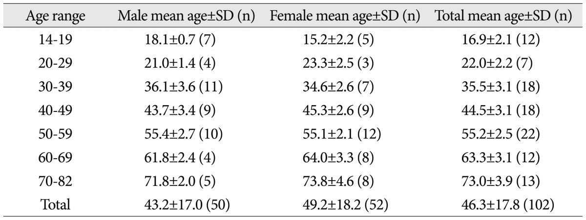
Asymptomatic subject's mean age and standard deviation
Written informed consent was obtained from all subjects. The subjects were carefully screened using a modified low back pain questionnaire and history taking. Individuals were only enrolled if they had never experienced relevant pain in the low back area, hip joint, and knee joint, never undergone radiation at the lower extremities and never had any neurological deficits.
The absence of relevant symptoms in these areas was defined as never having seen a physician, physiotherapist, chiropractor, acupuncture, oriental herb medication, or other such health care professional, and never having missed workday due to these symptoms. The rationale for these criteria was the notion that episodes of transient back pain are very common and less likely to be recalled after spontaneous regression4). Cases in which hospitalization treatment was administered for trauma such as a traffic accident were also excluded, because trauma required hospitalization may have undetected lumbar diseases.
MR imaging methods
From T12 to S1, magnetic resonance (MR) scans were performed with a 3.0-Tesla imager (Achieva 3.0 T X-series, Philips Medical Systems, Netherlands) with a dedicated receive-only spine coil. The protocol included sagittal T2-weighted [2500/100 (repetition time msec/echo time msec)] turbo spin-echo imaging of the entire lumbar spine with the following sequence parameters : matrix, 724×256; field of view, 240 mm; section thickness, 4 mm; intersection gap, 0.4 mm; and echo train lengths of 16 for T2-weighted images (T2WI). Though an axial sequence is also necessary to differentiated differences in disc pathology, but the authors use only sagittal imaging for convenience.
All images were sent to the in-hospital picture archive and communication system (PACS; Infinitt PACS, invented by Infinitt Co., Seoul, Korea) and reviewed using a 21.3-inch, 5-megapixel medical flat grayscale display (MFGD 5421, Barco, Tortrijk, Belgium) on a HP xw4400 workstation base unit (Hewlett-Packard Co., Palo Alto, CA, USA). The monitor has a 2048×2560 resolution, 0.165 mm pixel pitch, 422.4×337.9 mm active screen area, 800 : 1 dark room contrast, and 700 cd/m2 luminance.
Image analysis
The imaging studies in all 102 asymptomatic subjects were read independently by three of the authors (two neurosurgeons and one neuroradiologist) who were not given any information about the subjects. To eliminate potential reading bias, the images of 30 random symptomatic patients were mixed with the subject's images. We did not analyze the random 30 patients' results in final analysis. There was no case of blocked vertebra. Six lumbar disc levels were examined in each subject, and a total of 612 discs were examined.
Degree of degeneration
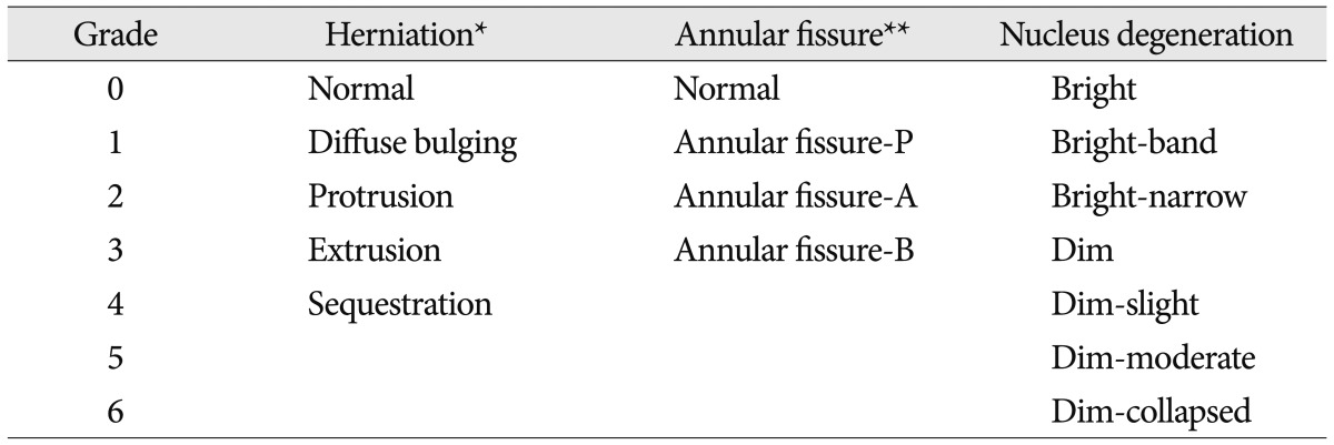
To establish the criteria for determining disc abnormality, we classified the three measurement categories : herniation (HN), annular fissure (AF), and nucleus degeneration (ND). There are many classifications for disc herniation and degeneration, we used the most prevalent terms7) for HN and modified Pfirrmann's classification15) for ND. The reason for the selection of the three categories is that they are abnormal findings with a high prevalence in normal healthy subjects.
Herniation (HN)
Disc herniation was classified as local protrusion in the intervertebral space. The following terms were used to describe disc abnormalities : normal, bulging, protrusion, extrusion, and sequestration7,16,22). Disc was considered to be normal when it did not reach beyond the borders of the adjacent vertebral bodies. Bulging was defined as a circumferential, symmetric disc extension around the vertebral border. Protrusion was defined as a focal or asymmetric extension of the disc beyond the vertebral border, with the disc origin broader than any other dimension of the protrusion. Extrusion was defined as a more extreme extension of the disc beyond the vertebral border, with the base against the disc of origin narrower than the diameter of the extruding material and a connection between the material and the disc of origin. Sequestration was defined as a free disc fragment that was distinct from the parent disc (Table 2, Fig. 1).
Degree of disc degeneration
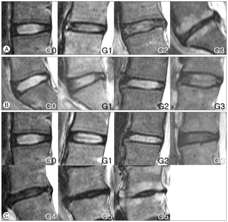
References of image for disc degeneration are shown. The criteria of classification are described in Table 2 and 3. A : Herniation. G0, normal. G1, diffuse bulging. G2, protrusion. G3, extrusion. B : Annular fissure. G0, normal. G1, annular fissure at posterior or posterolateral site. G2, annular fissure at anterior site. G3, annular fissure at anterior and posterior site. C : Nucleus degeneration. G0, normal. G1, normal disc height and hyperintense nucleus signal with horizontal dark band. G2, decreased disc height and hyperintense nucleus signal with or without horizontal dark band. G3, normal disc height and decreased nucleus signal with slightly or heterogeneous irregularity. G4, slightly decreased disc height and decreased nucleus signal with slightly or heterogeneous irregularity. G5, moderately decreased disc height and decreased nucleus signal with moderately. G6, collapsed disc height and hypointense nucleus signal.
Annular fissure (AF) and high-signal intensity zone (HIZ)
AF was defined as a high signal at the fibrous ring of an intervertebral disc on T2WI. In addition, high-signal intensity zone (HIZ) is a high-intensity signal (bright white) located in the substance of the annulus fibrosus, clearly dissociated from the signal of the nucleus pulposus. It is surrounded superiorly, inferiorly, posteriorly and anteriorly by the low-intensity (black) signal of the annulus fibrosus and is appreciably brighter than that of the nucleus pulposus. HIZ probably corresponds to annular tear1). AF was graded according to fissure site of the disc at the most clearly lesion from entire disc midsagittal T2WI. And the prevalence of HIZ was analyzed (Table 2, Fig. 1).
Nucleus degeneration (ND)
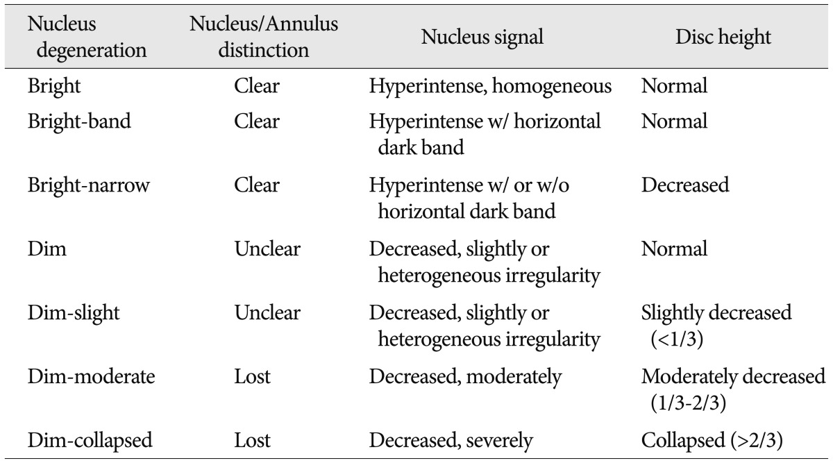
The extent of intervertebral disc ND was graded on midsagittal T2WI according to the criteria of modified Pfirrmann classification15). This grading correlates with the extent and loss of signal intensity in the nucleus pulposus of the intervertebral disc. Intervertebral disc narrowing was diagnosed in cases in which decrease of more than 33.3% in the intervertebral disc space was present. The seven findings in the lumbosacral intervertebral disc were : bright (homogeneous hyperintense nucleus), bright-band (nucleus clear distinction with horizontal dark band), bright-narrow (nucleus clear with disc height decreased), dim (nucleus unclear distinction with normal height), dim-slight (nucleus unclear with slightly decreased), dim-moderate (nucleus unclear with moderately decreased), dim-collapsed (nucleus unclear with collapsed). For the purpose of this investigation, grade 0-2 were grouped together and grade 3~6 were the more advanced grades of ND (Table 2, 3, Fig. 1).
Degree of nucleus degeneration
Analysis of the prevalence of abnormal findings
The prevalence of the various abnormalities was calculated by disc count (DC) and person count (PC). DC is the number of discs regardless of subjects (0-612) and PC is the number of subjects with disc degeneration (0-102).
Statistical analysis
The prevalence of the various abnormalities was calculated by averaging the scores of three readers. Inter-observer reproducibility was assessed using Kendall's coefficient of concordance. The Kendall correlation can be interpreted as the coefficient of concordance to measure the agreement among raters (0, no agreement; 1, complete agreement).
RESULTS
Degree of degeneration
Herniation (HN)
Disc bulging was observed in 103.0 (16.8%) of the 612 discs analyzed and in 61.3 (60.1%) of the 102 subjects (Table 4). This showed that an age-related proportional tendency was observed at the level of L1/2-L3/4. Marked increasing is observed at the level of T12/L1 in the 70's, L1/2 in the 60's, L2/3 in the 50's, L3/4 in the 40's, and L4/5 in the 30's. Even in teenagers, a high rate of herniation was observed at the level of L5/S1 (Fig. 2).
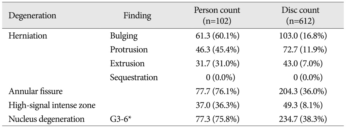
Abnormal disc degeneration findings according to person and disc count
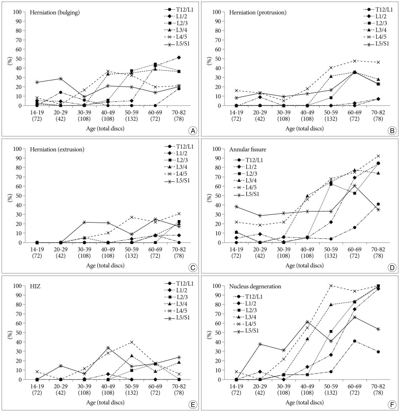
Graph of disc degeneration prevalence according to age and level, calculated by disc count. A, B and C : Herniation. D : Annular fissure. E : High-signal intensity zone (HIZ). F : Nucleus degeneration.
The authors found protrusion of disc in 72.7 (11.9%) of the 612 discs analyzed and in 46.3 (45.4%) of the 102 subjects (Table 4). This showed that a peak point was reached at the levels of T12/L1 and L1/2 in subjects aged 70 or older. However, in subjects aged 60 or older, it reached the level of L2/3-L5/S1 (Fig. 2).
Extrusion of disc was found in 43.0 (7.0%) of the 612 discs analyzed and in 31.7 (31.0%) of the 102 subjects (Table 4). This showed that L5/S1 level was high in the subjects aged 30 or older. L4/5 showed an age-related proportional tendency in the subjects aged 30 or older. There were no sequestrations of disc (Fig. 2).
Annular fissure (AF) and high-signal intensity zone (HIZ)
The authors found AF in 204.3 (36.0%) of the 612 discs analyzed and in 77.7 (76.1%) of the 102 subjects (Table 4). This demonstrated a marked increase at the level of T12/L1 in patients in their 60's and 70's. At the level of L1/2-L4/5, the graph was proportional to age. A high rate of AF was shown at the level of L4/5-L5/S1, even in teenagers, and an age-dependent slope was observed (Fig. 2).
HIZ of the disc was found in 49.3 (8.1%) of the 612 discs analyzed and in 37.0 (36.3%) of the 102 subjects (Table 4). HIZ was almost not seen at the level of T12/L1, L1/2. However, a peak value was found at the level of L2/3 and L3/4 in the 50's, and at the level of L5/S1 in the 40's. HIZ did not show increasing pattern in 60's and 70's (Fig. 2).
Nucleus degeneration (ND)
For the analysis of prevalence, grade 0-2 were grouped together and grade 3-6 were the more advanced grades of ND. We defined that advanced grades of ND is abnormal finding of disc. The authors found disc ND in 234.7 (38.3%) of the 612 discs analyzed and in 77.3 (75.8%) of the 102 subjects (Table 4). The graph of ND according to age and level was analyzed. The analysis showed a marked peak at the level of T12/L1 in the 60's. At the level of L1/2-L4/5, the graph was proportional to age. At the level of L5/S1, a high value was shown, even in the 20's, and an age-dependent smooth slope was observed (Fig. 2).
The results are summarized in Table 4. Table 4 is DC and PC of herniation, annular fissure, and nucleus degeneration of abnormal findings according to subject age and level.
Inter-observer reproducibility
The agreement values for the ND, AF, and HN were 0.973, 0.977, and 0.977 (p<0.0001), using Kendall's coefficient of concordance.
DISCUSSION
In the 1980s, when MRI was first introduced as a novel imaging modality, Modic et al.24) and Brown et al.8) reported that the accuracy of MRI was similar to or somewhat lower than that of CT or myelography. Furthermore, several authors have reported that the sensitivity of MRI in making a diagnosis of herniation and stenosis is much higher than that of compound diagnostic modalities using CT, myelography, and discography14,21,22,26). In particular, with the recently universalized use of 3 T MRI, the overall resolution power has improved due to the high magnitude of the magnetic field as well as surface coil design and computer programs. The major advantage of 3 T MRI is the marked increase in spatial resolution for both 2D and 3D techniques, afforded by the increase in signal-to-noise ratio. The value of 3 T MRI of the spine is in the greater achievable spatial resolution, which facilitates imaging with thinner slices and leads to markedly improved depiction of small structures such as the spinal cord, nerve roots, and neural foramina. Thus it is possible to routinely acquire 2 mm slice sections in sagittal imaging of the spine at 3 T compared with 3 to 4 mm slices at 1.5 T13).
There are some controversies regarding the correlations between abnormal MRI findings and lumbar pain. MRI can visualize pathologic changes and detailed anatomical structures, but it cannot show the clinical symptoms in many cases. Back pain could have originated from degenerative causes decreasing the disc height, end plate change and facet joint degeneration20,23). Many studies have suggested that AF and HIZ were more predictive of discogenic pain1,27). However, HIZ were also frequently observed in asymptomatic subjects28). Abnormal findings of herniation may be more relevant when nerve root compromise can be demonstrated4). Therefore, the range and prevalence of asymptomatic structural abnormalities must be elucidated in order to assess the importance of abnormal findings on MRI. Several studies have shown that the abnormal finding rates of myelography and CT reached a high value of 24-37% in a number of individuals without back pain17,18,31,32). In 1990, Boden et al.3) showed that the abnormal finding rate of cervical MRI amounted to a value of 28% in subjects aged 50 or older, and lumbar MRI amounted to 57% in subjects aged 60 or older2). These results have caused us to question our current image-based understanding of the pathogenesis of back pain.
Our analysis showed that the prevalence of HN, AF, and ND were 81.4%, 76.1%, and 75.8%, respectively, in asymptomatic individuals. Despite the lack of a comparison between our results and those reported in previous literatures, abnormal findings on MRI have been observed at a high prevalence of more than 75-80%. According to a review of the literature, the prevalence of protrusion, extrusion, HIZ, and ND in symptomatic cases were 54.4%, 34.8%, 59%, and 95.7%, respectively4,10). Our results showed high prevalence as in symptomatic cases. These results lead us to reconsider the relationships between abnormal findings on MRI and symptoms.
We reviewed literatures about MR images in asymptomatic subjects (Table 5). The most different method was not using open call recruitment. We stayed all day long at doctor's office of Health Screening Center and choose subject by history-taking without mention of study purpose. The overall results in the table were high prevalence in extrusion, AF, and ND.

International literatures comparison of herniation, annular fissure, and nucleus degeneration in asymptomatic subjects (%)
Because all studies used different criteria, we performed a modified statistical analysis (Table 6). This comparison was made in the same manner as the previous reports, using the previous criteria of grading and selecting age of population. The modified results were similar to the original results. However, the rates of bulging and protrusion were slightly lower than the authors' original results. The modified rate of DC may have been 2-4% lower than the original result because normal discs were included in the results of DC (despite a lack of effect on PC), as the authors' study included the spinal level of T12/L1.
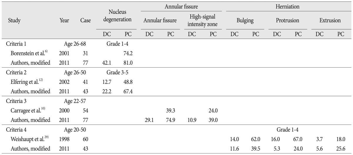
Comparison of herniation, annular fissure, and nucleus degeneration between the previous reports and authors' modified results which were changed according to the criteria of previous studies
The author's results showed high prevalence as previous literatures. This can be explained by structural and personal factors. The structural factor is the improvement of resolution with 3 T MRI and subdivided degeneration grading, and the personal factor is the anthropologic character and cross-legged sitting life style. We use a more detailed grading system applied to not symptomatic patient but asymptomatic subject. And we use 3 T MRI. But, even though the overall resolution power of MRI improved in spatial resolution by the 3 T MRI, the authors thought that 3 T MRI might not have far superior images in spine than 1.5 T MRI. MRI factor would have a small effect on high prevalence results. Population-specific prevalence would play a great role as well as the MRI field strength and the modified grading systems. Further research is necessary to compare the findings of 3 T to 1.5 T MRIs in the same Korean subjects.
The limitation of our study is the lack of axial images. But, we examined the whole sequential sagittal images. This makes the definition of bulging versus protrusion difficult. So, bulging and protrusion would show lower prevalence than previous literatures, unlike ND and AF. If we get axial images, bulging and protrusion would show higher prevalence.
Nevertheless, high prevalence of degeneration in asymptomatic subjects 10-29 years of age is very interesting. Multiple studies have shown similar findings that asymptomatic patients are known to have a high prevalence of disc abnormalities, especially in advanced age. We found close to 25% of 10-19 years of age have bulging discs at L5/S1 (Fig. 2). This increases to nearly 30% from ages 20-29. In protrusion, we found close to 15% of asymptomatic subjects age 10-19 and 20-29 at L4/5. Finally, the total prevalence of bulging and protrusion is close to 17% at L4/5 and 33% at L5/S1 in ages 10-19. And this increases to nearly 15% at L4/5 and 45% at L5/S1 in ages 20-29. In ND, we found close to 38% of asymptomatic subjects age 20-29 at L5/S1, while nearly normal was shown in ages 10-19. The authors thought that low grade degeneration would not be pathologic in children and adolescents.
Another limitation may be found in the selection of subjects which was done from asymptomatic visitors for health screening rather than from the normal population. All previous studies had also limited conditions, such as open volunteer recruitment. It is very difficult and nearly impossible to make a full comprehensive evaluation for disc degeneration.
The cause of the high prevalence of disc abnormal finding may be from Korean Ondol life style of cross-legged sitting, agricultural society, and anthropological factors. And, we expect different results if our study is repeated after several decades, because most people will have chair life style, instead of cross-legged life style. The authors believe that this study would have educational worth for asymptomatic degenerative findings in lumbar disc.
CONCLUSION
In asymptomatic Korean subjects, the abnormal findings of MRI showed high prevalence of annular fissure, nucleus degeneration, and advanced grade disc herniation (extrusion). Especially in young ages, the authors found that bulging, protrusion, and annular fissure showed high prevalence at L4/5 and L5/S1. And nuclear degeneration showed high prevalence at L5/S1. Therefore, all lumbar disc degenerations are not pathologic, especially in children and adolescents.
Acknowledgements
The authors thank Ji Min Sung, Ph.D. and Dae Hyung Lee, Ph.D., for their assistance in statistical design and testing.