INTRODUCTION
The iliac crest autograft has long been considered as a gold standard in achieving in surgical spinal fusion [13]. However, the numerous disadvantages such as graft site morbidity, increased operative time, increased blood loss, risk of neurovascular injury, increased duration of hospital stay and cosmetic concerns are associated with autologous iliac crest grafting [4]. Apart from this, the fact remains that the supply of iliac crest bone graft in a patient is finite and may not be sufficient in every case. Thus, there has been a constant quest to find substitutes to autologous bone grafting [24].
Allografts, demineralized bone matrix (DBM), ceramics and recombinant bone morphogenic protein-2 (rhBMP-2) are some of the substitutes that have been investigated to replace autografts for achieving interbody fusion, with varying success rates [8,9,11,16]. The ABM/P-15 (anorganic bone matrix/15-amino acid peptide fragment) is a synthetically manufactured P-15 collagen peptide fragment, that is adsorbed on ABM and suspended in an inert hydroge [l1]. This P-15 imitates the cell-binding domain of type one collagen and triggers biomechanical signals which ultimately result in new bone formation [6,12]. The use of ABM/P-15 has been well established in single level anterior cervical fusion surgeries [2]. However, in the cases of lumbar fusion surgeries, more evidence is needed to establish its efficiency and safety. To address this lacuna in current scientific evidence, we compared ABM/P-15 with other commonly used bone graft substitute materials in lumbar interbody fusion surgeries namely rhBMP-2, and DBM in terms of their clinical and radiological outcomes as well as the side effect profile.
MATERIALS AND METHODS
Institutional Review Board (IRB) of Wooridul Spine Hospital approval was taken for this study (IRB No. 2021/04/WSH/002). A retrospective analysis of prospectively collected data of 140 patients with degenerative lumbar spinal pathologies, who underwent instrumented lumbar interbody fusion surgeries in a single speciality spine centre between the years 2016 and 2020 was conducted. The indications for surgery were lumbar degenerative disorders with instability/spondylolisthesis wherein fusion was deemed necessary. All patients with a minimum follow-up of 6 months were included. Patients with inadequate follow-ups, missed timely follow-ups, inadequate documentation of the records were excluded. Patients with concomitant posterior decompression at the index level, traumatic pathologies, infections, inflammatory or autoimmune diseases and tumours were excluded. The patients were operated either using anterior lumbar interbody fusion (ALIF), or oblique lumbar interbody fusion (OLIF)/direct lateral lumbar interbody fusion (DLIF) approach followed by percutaneous posterior pedicle screw fixation. The patients were divided into three groups based on the bone graft substitute used, namely ABM/P-15, rhBMP-2, and DBM group. The use of biologic grafts was applied consecutively according to the time of surgery; DBM was used from the year 2016 to August 2017, BMP was used from September 2017 to August 2019, and ABM/P-15 was used for surgery after September 2019. Following are the specifications and doses of the substitutes used : 1) rhBMP-2 (Novosis; CGBio, Seoul, Korea) : rhBMP-2 + HA carrier (0.5 mg/level) mixed with allograft bone, 2) ABM/P-15 (i-Factor; Cerapedics, Westminster, CO, USA) : 1 mL/level mixed with allograft, and 3) DBM (Grafton Orthoblend; Medtronic, Memphis, TN, USA) : 5 mL/level (allograft mixed formula).
Commercially available cancellous allograft bone derived from the femoral head was used.
Clinical evaluation
All the patients were evaluated pre-operatively as well as in post-operative follow-up period using 10-point Visual analogue scale (VAS) for back pain and leg pain, and Oswestry disability index (ODI) was calculated in each of the patients. The incidences of complication such as infection, hematoma, wound complication, implant failure as well as unplanned revision and readmission were also noted.
Radiological evaluation
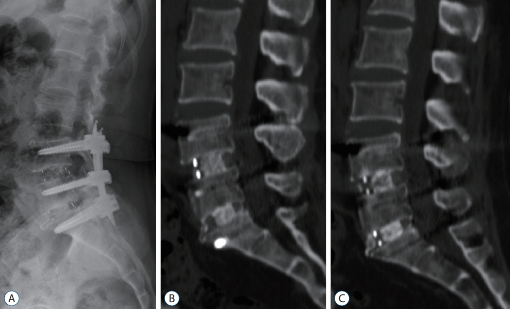
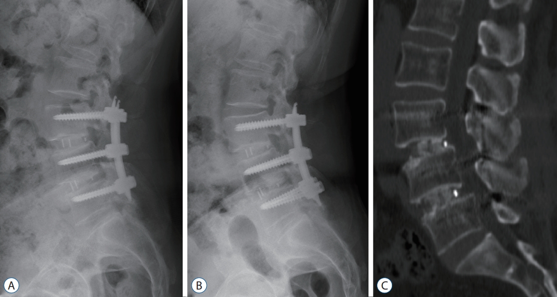
The patients underwent full-length, 36-inch exposure radiographs of the spine that extended from the base of the skull to the proximal femur in the antero-posterior and lateral planes pre-operatively, post-operatively in first week and on regular follow-up visits of 3, 6, 9, and 12 months postoperatively. All radiographs were obtained with the patients standing and looking forward trying to maintain a horizontal gaze and with their arms flexed, hands placed on their clavicles without any support, and knees extended. Radiological parameters such as pelvic incidence, sacral slope, lumbar lordosis, segmental Cobb’s angle, sagittal vertical axis, and cage subsidence were calculated in each radiograph using a program that included a built-in picture-archiving communication system (PiView; INFINITT Co. Ltd, Seoul, Korea). Lumbar dynamic radiographs were taken at each follow-up visit while computed tomography (CT) scan was done at 3-month and 12-month follow-up visit. This is done as an institutional protocol in our hospital as more frequent CT scans (which although have more diagnostic accuracy for detecting fusion) may result in radiation hazard to the patient. Thus, the interbody fusion was assessed on CT reconstruction images and/or flexion-extension lateral radiographs at the abovementioned follow-up intervals. Bone fusion was defined as solid when there was osseous continuity observed in CT reconstruction images and mobility of less than 4° on as seen in flexion-extension lateral radiographs. Nonunion was defined as the presence of a visible gap and mobility greater than 4° [19].
Statistical analysis
The three groups were analysed for various clinical and radiological parameters using two-way analysis of variance (ANOVA), one-way ANOVA, chi-square test/fisher exact test. A subgroup analysis of rhBMP-2 and ABM/P-15 was also done to compare these two modalities. A p-value of <0.05 was considered as statistically significant. All analyses were performed using SPSS 14.0K (SPSS Inc., Chicago, IL, USA).
RESULTS
Overall, 401 patients (116 ABM/P-15, 108 rhBMP-2, and 177 DBM) were identified to have undergone anterior or lateral lumbar interbody fusion surgery between the study time period. After applying the inclusion criteria, 140 patients were enrolled for the study. Out of 140 patients, ABM/P-15, rhBMP-2, and DBM were used in 46, 44, and 50 patients, respectively. Average age of the entire study population was 67.15 years with 88.43% female patients. Patient characteristics like age, gender, bone mineral density smoking history and presence of diabetes mellitus were comparable amongst three groups (Table 1).
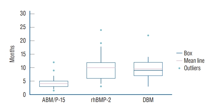
Average follow-up was 16.0±5.2, 17.9±9.8, and 26.2±14.9 months, respectively in ABM/P-15, rhBMP-2, and DBM groups. Majority of the patients underwent ALIF surgery in ABM/P-15 (82.8%), rhBMP-2 (51.2%), and DBM (84%) groups. The average time to union was 4.05±2.01, 10.00±4.28, and 9.44±3.49 months, respectively for ABM/P-15, rhBMP-2, and DBM groups (p<0.001) (Fig. 1). At the last follow-up, fusion was achieved 97.9%, 93.2%, and 98% in ABM/P-15, rhBMP-2, and DBM groups, respectively. Eighty-nine percent patients treated using ABM/P-15 showed fusion within first 6 months (Table 2 and Figs. 2-4).
The average pre-operative back VAS score was 6.93±2.42, 7.14±1.97, 7.01±2.14 (p=0.900), respectively, which reduced to 1.02±0.80, 1.21±0.96, and 0.54±0.70 (p=0.112) at the last follow-up. The pre-operative ODI scores were 52.70±18.02, 55.39 ±16.80, and 53.56±19.61 (p=0.751), which in the post-operative period reduced to 33.77±15.52, 39.42±16.47, and 38.30±15.89 (p=0.412), respectively and to 15.74±8.30, 17.41±10.45, and 16.76±9.81 (p=0.603), respectively at the last follow-up (Table 3).
There were no incidences of infection in ABM/P-15 and rhBMP-2 group while three patients in DBM group were infected. One patient in each rhBMP-2 and ABM/P-15 group, while two patients in DBM group suffered superficial wound complications. The incidence of cage subsidence was 21.7%, 30.2%, and 14%, respectively in ABM/P-15, rh-BMP-2, and DBM group (p=0.332). Most of this subsidence was grade 1 (76.9%) while 15.4% of the subsidence was grade 2 (80% of the subsidence in ABM/P-15 group, 72% in rh-BMP-2 group, and 85.7% in DBM group was grade 1 subsidence).
DISCUSSION
Lumbar interbody fusions are one of the commonest spine surgeries performed in the world and the incidence of this procedure is rising steadily [22]. Achieving predictable and rapid fusion is necessary for a successful outcome in these patients. The use of bone graft substitutes has been prompted by the numerous disadvantages in the use of autologous iliac crest bone graft, most notably post-operative pain at the donor site [29]. There has been a constant effort to develop an ideal bone graft substitute which would possess all the three properties of an autologous bone graft, namely osteogenesis, osteoinduction, and osteoconduction. However, none of the synthetically derived materials have shown to possess all the three properties [31]. Nevertheless, many of these substances have shown clinical usefulness in achieving fusion. In this study we compared three commonly used bone graft substitutes, which have gathered attention in recent years, namely ABM/P-15, rhBMP-2, and DBM.
DBM, which is created by demineralization of human cadaveric bones using acid, has osteoinductive as well as osteoconductive properties and has shown fusion rates of approximately 90% [25]. DBM contains multitudes of proteins such as low molecular weight BMPs, collagen and some non-collagenous materials which have pivotal roles in bone formation [15].
rhBMP-2 has a higher osteoinductive as well as osteoconductive properties than DBM, however it has shown varying fusion rates ranging from 40-100% [25]. Another point of concern with rhBMP-2 has been the risk of heterotrophic ossification, osteolysis, subsidence, inflammation and neoplasm [25]. It has been reported that the actual rate of complications after the use of rhBMP-2 is 10 to 50 times higher than what has been reported by the industry sponsored clinical trials [7]. In our study, we could not find evidence of ectopic bone formation or neoplastic change in any of the three groups.
The earlier evidences and consequently the clinical use of ABM/P-15 was established in various orthopaedic as well as dental surgeries [5,10,27] whereby the ABM/P-15 was used in the treatments of long bone non-unions as well as in gingival recession of defects and periodontal defects. Extrapolating the successful experience from these subspecialities, the use of ABM/P-15 was investigated in spinal surgery.
Arnold et al. [2] conducted a randomised controlled trial to investigate the non-inferiority of ABM/P-15 bone graft as compared to autologous bone graft in single level cervical fusion surgery for cervical radiculopathy. ABM/P-15 and autologous graft group showed 88.97% and 85.82%, fusion rates respectively at the end of 1-year follow up. They concluded that ABM/P-15 meets the noninferiority success criteria as per the FDA requirements and it was safe as well as effective in single-level anterior cervical interbody fusion in cases of cervical radiculopathy.
There are very few studies in the literature investigating lumbar spinal fusion using ABM/P-15. In a study by Lauweryns and Raskin [18] the efficacy and safety of ABM/P-15 in 40 patients treated with posterior lumbar interbody fusion was analysed. In their study they inserted two cages at each level to be fused. One cage was filled with autologous graft while the other was filled with ABM/P-15. They found that the evidence of intra cage bone bridging at 6 months was seen in >97% cages with ABM/P-15 and, 59% cages with autologous graft. Mobbs et al. [25] studied the clinical outcomes and fusion rates after ALIF surgeries using ABM/P-15, and found that at a mean 2 years follow up, fusion was seen in 97.5%, 81%, and 100% of patients, who underwent single, double, and triplelevel fusion surgeries respectively.
On the other hand, Jacobsen et al. [14] conducted a randomised controlled trial comparing the fusion rates in elderly Scandinavian population treated with non-instrumented ALIF using allografts and ABM/P-15. They found that at 1 year post surgery, the rate of fusion in the ABM/P-15 group was 50% while that in the allograft group was 20%. All the patients in this study were elderly with osteoporosis and no instrumentation was used for fixing the spinal segments. Since rigid fixation and stability provided by the instrumentation has an important mechanical role in preventing instability and movement at the spinal segment [23], not using any instrumentation may be responsible for the relatively lower rates of fusion reported in this study.
In our study the rate of fusion in the ABM/P-15 group was 97.9%, with 89.1 % patients showing radiological evidence of fusion within 6-month post-surgery. The average time to union of 4.05 months in ABM/P-15 group which was significantly lower than the other two (10 months and 9.44 months respectively for rhBMP-2 and DBM group). Apart from this, there was no significant difference between the health-related quality of life (HRQoL) parameters measured by the ODI scores among the three groups. This highlights the efficiency of ABM/P-15 in achieving rapid fusion while achieving comparable HRQoL outcomes.
Cage subsidence was the most common complication in our study. We had as 30.2% subsidence rate in rhBMP-2 group followed by ABM/P-15 group and DBM group with 21.7% and 14% subsidence rate. However, this difference was not statistically significant when compared between all the three groups. As per the classification proposed by Sharma et al. [30] most of these patients had grade 1 subsidence. The effect of subsidence on the overall clinical outcome was not unfavourable as suggested by the back and leg VAS scores and ODI scores. This was expected, as low-grade subsidence is considered an expected outcome, rather than a surgical complication [21,22]. The incidence of deep infection was 6% in DBM group, with no patients infected in ABM/P-15 and rhBMP-2 group. The superficial wound complications were seen in one patient each in rhBMP-2 and ABM/P-15 group and in two patients in DBM group.
Heterotrophic ossification has been reported as one of the side effects of using ABM/P-15 as described in a case report of an 8-year-old patient with mucopolysaccharidosis treated with spinal fusion using ABM/P-15 [28]. However, in our study we could not find evidence of heterotrophic ossification in any patient. This also has a bearing with surgical technique, wherein, it is advised not to perform saline irrigation of the wound once the cage with ABM/P-15 is inserted as it can lead to localised deposition of ABM/P-15 along with the irrigation fluid which may stimulate ossification at ectopic sites. In addition to this, the authors seal the cage using either oxidized regenerated cellulose (Surgicel®; Johnson and Johnson Medical, Arlington, TX, USA) or a water-soluble bone hemostatic agent (Ostene, Baxer, CA, USA) to prevent the ABM/P-15 from flowing out of the cage.
Our study is unique in many aspects. In this study, we have compared three types of bone graft substitutes namely ABM/P-15, rhBMP-2, and DBM and have compared their clinical and radiological outcomes as well as complication profile. Apart from this, the study also encompasses cases treated with ALIF, OLIF, and DLIF approaches, followed by percutaneous pedicle screw fixation which have recently gathered more attention as minimally invasive fusion approaches [3,17,20,26,32]. Limitations of our study include the retrospective design and shorter follow-up. Thus, some of the complications like the heterotrophic ossification may not become apparent in the short-term follow-up. As the bone graft substitutes were used serially over a period of 5 years it may be possible that the experience of the providing surgeon(s) increased which may theoretically improve patient selection, risk mitigation, and surgical technical skill, which can impact the outcomes. At present, a prospective randomised controlled double blinded multicentric trial is planned to obtain high quality, level one evidence to further investigate the efficacy as well as cost effectiveness of ABM/P-15 in lumbar spinal fusion.
CONCLUSION
ABM/P-15 appears to achieve union in lumbar fusion cases significantly earlier than rhBMP-2 and DBM, while maintaining favorable clinical and complication profile. Further prospective long term clinical trials comparing different bone graft substitutes with emphasis on the cost analysis should be undertaken to further build on the currently available evidence.