INTRODUCTION
Since the brain is encased in the unyielding skull vault, any increase in its volume will increase the intracranial pressure (ICP) and result in critical outcomes. Decompressive craniectomy (DC) can partially remove the unyielding skull vault and make an affordable space for the expansion of swelling brain contents. The limited brain space is increased after the DC. This might decrease ICP, thus improving the cerebral perfusion pressure (CPP) [2-4,19,25,26,30,33,39,41]. Increased ICP is a major predictor of poor prognosis or death of patients with traumatic brain injury (TBI) [5,6,11,19,25,28,30,33,34,36,37]. Despite the highly developed medical treatment options, DC might be the last and very effective treatment option for patients with intractable increase of ICP [1,8-11,13,33].
Many previous studies have reported benefits and limitations of DC in TBI [2,3,6,10,11,15,20,30,33,37], hypertensive intracerebral hemorrhage (HICH) [4,12,15,40], major infarction (MI) [13,15,17,21,23,32,35,42], and aneurysmal subarachnoid hemorrhage (SAH) [15,35]. These reports were concerned with the DC. They were performed using various DC surgical methods, such as bifrontal craniectomy, unilateral or bilateral hemi-craniectomy, suboccipital craniectomy, and non-specific craniectomy with various sizes. Several reports have compared DC effects according to causative diseases, with various results according to the study [15,37]. Accumulating data have proven that DC is effective in ICP treatment for improving cerebral perfusion and neurologic outcomes [2,3,6,10,12,13,18,25,26,30,33,37].
Recent papers recommend larger DC area is essential for improve the neurologic outcomes [8]. However, it is very rare that has compared DC effects according to surgical methods. These authors usually prefer DC for the last step in treating massive brain swelling patients. Sometimes surgical method, unilateral or bilateral DC, is also a variable and also DC area is a variable. In this study, clinical effects were compared with Glasgow outcome scale (GOS; at 6 months after DC), ICP control effect (initial ICP-postoperative mean ICP), and midline shift improvement (midline shift preoperative-24 hours after operation) according to decompressive bone size. Decompressive bone size (total DC area and major side DC area) was also compared between unilateral and bilateral DC.
MATERIALS AND METHODS
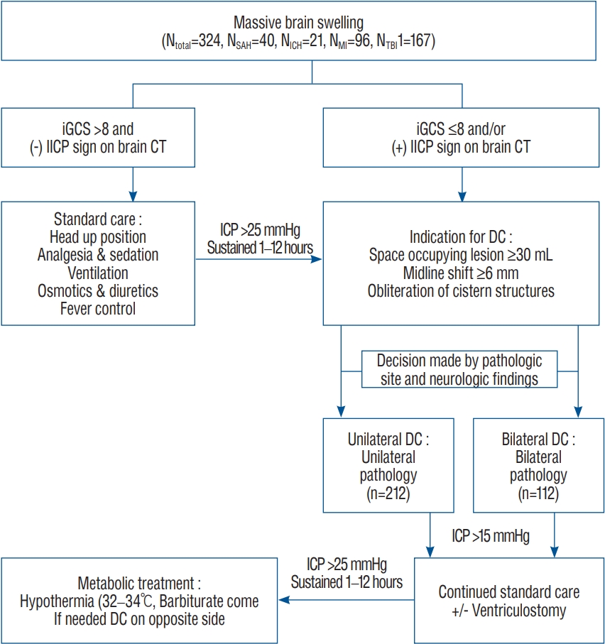
The treatment protocol (Fig. 1) was approved by the Institutional Review Board of Eunpyeong St. Mary's Hospital (PC17RES10027). All patients or their representatives provided informed written consent for surgical management. This study was a retrospective study through observatory data analysis.
Patient population
Between September 2012 and August 2019, patients underwent DC. They were monitored for ICP during and after the surgery. In these patients, initial ICP, postoperative ICP, mean arterial blood pressure, CPP, and decompression bone size were measured. A total of 324 patients who underwent unilateral DC (212 patients) or bilateral DC (112 patients) were included in this retrospective analysis, consisting of aneurysmal SAH (40 cases), massive intracranial hemorrhage (ICH; 21 cases), MI (96 cases), and TBI (167 cases). Seventeen patients underwent multiple DC operation (one patient three times). They were not included in this study. The mean age of all patients was 53.4 years (median, 54 years; male : female, 225 : 99). Standard image study, included computed tomography (CT) scanning undertook as soon as the patient arrival in the emergency room or showing neurological deterioration (decrease Glasgow coma scale [GCS] more than 2 grade or National Institute of Health Stroke Scale more than score 4). All these preoperative brain CT were performed within three hours prior to DC surgery.
Indications for surgery
Indications for decompressive surgery included appearance of definite brain swelling on CT scan as indicated by a midline shift of more than 6 mm and/or obliteration of the cistern structures and space occupying lesion more than 30 mL. Clinical deterioration was defined as worsening neurological status (GCS score of less than 8 or rapid decreasing GCS score of more than 2 points). Patients who showed brainstem failure without spontaneous respirations did not undergo DC surgery [23,24,27]. Surgical methods (unilateral or bilateral hemicraniectomy) and DC area were decided by pathologic site and neurologic findings (Fig. 2).
Operation procedures
The operation was carried out under general endotracheal anesthesia for all patients. Patient was placed in a supine position. For patients undergoing bilateral hemi-craniectomy, the head was positioned straight and somewhat extended on the head-rest so that the occipital area is in most dependent position. For patients undergoing unilateral hemi-craniectomy, the head was turned to the opposite side as far as the operation side. The ventricular puncture was performed at Kocher’s point on the opposite side of the offending lesion or right side for the cases with no definite offending lesion. For some cases, a navigation system was used for accurate ventricular puncture. An extraventricular drainage (EVD) tube (EVD catheter; Yushin Medical, Seoul, Korea) was connected to the continuous monitor (CPP-monitor; Spiegelberg, Hamburg, Germany) via transducer device (Druckmeß-set; Smiths industries, Bayern, Germany). Large bicoronal skin flap was made for bilateral DC patients while large question mark flap was made for unilateral DC patients. Their limbs were placed behind the parietal eminence, extending inferiorly to the zygoma on both or operation sides. The scalp was then dissected sub-periostealy to the level of supraorbital ridges. After the ventricular ICP was stabilized, 2 or 3 burr holes were connected with a pneumatic saw, with subsequent removal of bone flap. In bilateral DC patients, the frontal median segment of the bone measuring about 3-4 cm in width along the sagittal sinus was saved in order not to damage the sagittal sinus and to function as a framework for later cranioplasty. Additional bone was removed at the temporal region to the floor of the middle fossa (Fig. 1). The dura was then opened in a large cruciated or curved Z shaped incision in areas involving frontal, temporal, and parietal lobes. When the dura was opened, underlying brain or hematoma typically herniated outward. Cortical resection of the brain was not performed. In all DC patients, artificial dura (Lyoplant; B. Braun Melsungen AG, Melsungen, Germany) was placed underneath the incised dura to allow the brain to herniate outward in a more controlled manner and to prevent cortical adhesion. After that, several pieces of gel-forms were inserted between the dura and muscle layer to control postoperative bleeding and allow easy dissection for cranioplasty afterward. Temporalis muscle and skin flap were then reapproximated with sutures. The bone flap was typically maintained in wet gauze at -70°C until reinsertion at 6-12 weeks after the initial surgery.
Postoperative management
If the ventricular pressure exceeded 15 mmHg after decompressive surgery, conventional medical management methods including the use of hyperosmotic agents and EVD were initiated. If the ventricular pressure exceeded 25 mmHg, regardless of medical therapy, either hypothermia (rectal temperature, 32-34°C) or coma therapy was initiated with a cold blanket and/or barbiturate.
Determination of decompression bone size
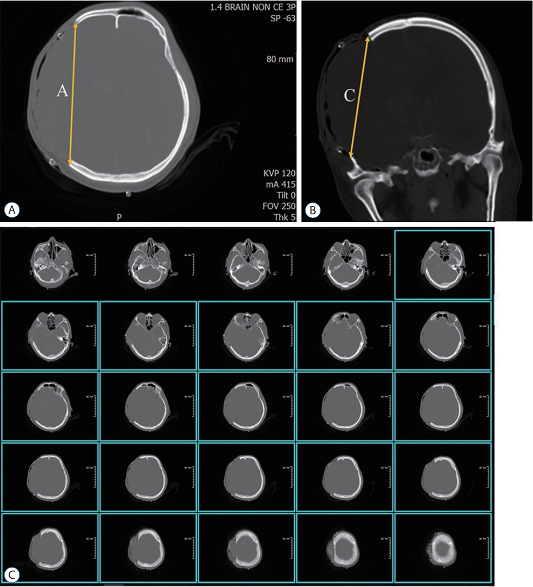
DC area was determined by directly multiplying long and short diameters of bone fragments in the operating room with an autoclaved ruler. If sizing of the bone fragment was missed in the operating room, 3D CT reconstruction image was undertaken to measure long and short diameters of the skull bone defect area. DC area was calculated using the simple AC method [16]. A was the longest length of craniectomy on the axial slices (Fig. 2A), and C was the longest length of craniectomy on the coronal slice (Fig. 2B). If the coronal slice did not exist, C was calculated the number of axial cuts by craniectomy and slice thickness (Fig. 2C). Total DC area of bilateral DC was calculated sum of each DC area. In this study, total cranial vault surface area was estimated as half of the sphere surface area. The radius was defined as the mean of the half distance from glabella to inion plus half distance from external auditory canal to vertex. Total DC area was defined as the sum of both sides of DC area if it was a bilateral DC case. DC area percent was calculated as DC area divided by the total cranial vault surface area. Offending side DC surface removal rate (DC%) was defined as DC area divided by half of the cranial vault surface area in the pathologic side.
Data collection
Neurologic outcome was evaluated at 6 months after the initial surgery. Favorable outcome (GOS 4-5) and mortality rate were compared according to surgical methods (unilateral vs. bilateral DC) and DC area. The initial ICP reading was considered to be the highest sustained ventricular pressure for 5-10 minutes after ventricular puncture. Ventricular pressure values were collected using continuous monitoring during intraoperative and postoperative periods (data were analyzed between postoperative 2-5 days). The midline shift was defined as the distance from the septum pellucidum to the midline between the anterior (crista galli) and posterior (torcular herophili).
Statistical analysis
All data are presented as means±standard deviations and/or medians. A Wilcoxon signed-rank sum test was used to analyze GCS and GOS scores. Comparisons among groups were performed using unpaired t-test and Fisher’s exact test. Statistical analyses for each outcome were analyzed with SPSS software version 12 (SPSS Inc., Chicago, IL, USA). For all statistical analyses, significance was defined at p≤0.05.
RESULTS
Comparison of ICP before and after the DC surgery
DC surgery was effective for ICP change (32.3±16.7 mmHg vs. 19.2±13.4 mmHg, p<0.001) and midline shift change (12.5 ±7.6 mm vs. 7.8±6.9 mm, p<0.001) before and after DC surgery (Table 1).
Comparison of clinical results between unilateral and bilateral DC
Total DC area was larger in the bilateral DC group (198.2± 43.0 cm2 vs. 125.1±27.8 cm2, p<0.001). And the offending side DC area was larger in the unilateral group, although the difference was not statistically significant (113.6±25.9 cm2 vs. 125.1±27.8 cm2, p=0.753) (Table 1).
The initial GCS (p=0.001) and neurologic outcome (GOS, p=0.009) was better in the unilateral group. The initial ICP (p=0.807), postoperative ICP (p=0.994), and ICP change (initial ICP-postoperative ventricular ICP, p=0.441) were similar between unilateral and bilateral DC groups. Midline shift change (preoperative-postoperative midline shift) was only statistically significant in the bilateral DC group (p=0.015) (Table 1).
Correlations between clinical outcomes and DC area
Total DC area and total DC% failed to show significant correlations with neurologic outcome (GOS : p=0.170 and p=0.396, respectively). Total DC area was not correlated with ICP change (p=0.147). However, large total DC% was correlated well with ICP change (p=0.021). Total DC area and total DC% were correlated with midline shift improvement (p=0.003 and p=0.002, respectively).
Offending side DC area and offending side DC% failed to show significant correlations with neurologic outcome (GOS : p=0.805 and p=0.567). Offending side DC area and DC% failed to show significant correlation with ICP change either (p=0.195 and p=0.871), although they showed significant correlations with midline shift change.
Total DC area over 160 cm2 and DC% over 46%showed statistically better neurologic outcome (p=0.020 and p=0.037).
DISCUSSION
DC has been performed for uncontrollable brain swelling since the early 20th century. Kocher (1901) and Cushing (1905) might be the first ones who described surgical decompressive craniectomies for therapeutic purpose in neurosurgical history [22,25]. DC is very effective in reducing the elevated ICP caused by brain swelling. It is well associated with improved neurologic outcomes [2,3,11,13,23,30,33,37,39,41]. Many papers have reported that DC can be a lifesaving procedure in various neurosurgical disorders, including TBI [2,3,10,11,15,20,30,33,37], ICH [4,12,15,39], MI [13,15,17,21,23,32,38,42], SAH [15,35], and other brain pathologies [15,18,26].
Since the brain is encased in the unyielding skull vault, any increase in its volume makes the ICP to be above the physiological level. The therapeutic principle of DC is to remove a part of cranial vault and give brain tissue room to expand. Therefore, it increases the compliance which results in a rightshift of the pressure-volume curve. At last, DC can lower the ICP and improve CPP, cerebral blood flow, and brain tissue oxygenation [2-6,19,25,26,30,37,41].
Several surgical methods of DC have been defined depending on the location and size of bone removal [10,12,13,20,23,25,33]. Reviewing previous reports, bifrontal craniectomy, unilateral or bilateral hemicraniectomy, and suboccipital craniectomy might be the most common adopted surgical methods for cranial decompression [10,12,13,15,17,21,24,25,30,37,41]. Many studies have reported benefits and limitations of DC and compared surgical effects according to causative diseases [15,24,39]. Some reports insist that DC should be as large as possible or bone fragment to be removed should be at least 12×15 cm in size or have a diameter of 15 cm [8,29,31].
In our data included, those who underwent unilateral or bilateral hemicraniectomy for supra-tentorial pathologies included four disease entities : anterior circulation cerebral aneurysms, anterior circulation major cerebral infarction, HICH, and TBI. Until now, few studies have compared clinical effects according to DC size and surgical methods. In authors’ institute, surgical indications for decompressive operation and surgical techniques were constantly applied. However, the decision to perform either unilateral or bilateral DC, the amount of DC area was made by the operating surgeon. We analyzed clinical outcomes, ICP change, and other clinical factors according to surgical methods and DC area.
In our analysis, total DC area and DC% were larger in the bilateral DC group than those in the unilateral group (both p<0.001, Table 1). However, offending side DC area and offending side DC% failed to show statistically significant difference in either DC group (p=0.753 and p=0.669). DC surgery was effective for ICP change (32.3±16.7 mmHg vs. 19.2± 13.4 mmHg, p<0.001) and midline shift change (12.5±7.6 mm vs. 7.8±6.9 mm, p<0.001). In the bilateral DC group, total DC area was larger (125.1±27.8 cm2 in the unilateral DC group vs. 198.2±43.0 cm2 in the bilateral group, p<0.001). And bilateral DC group shows more midline shift change (p=0.015) than the unilateral group. This favorable midline shift change in bilateral DC group was not correlated with clinical outcomes, so midline shift improvement was not correlated with ICP decreasement (p=0.441) (Table 1).
In some reports, larger DC is more effective than a standard DC in reducing the ICP, midline shift, and the incidence of secondary brain injury [3,20,25-27,29,31,39]. Recent guideline regarding TBI treatment recommends that DC area should be at least 12×15 cm (=180 cm2) to improve the neurologic outcome [8]. In the present study, total DC area over 160 cm2 showed significantly better neurologic outcome (p=0.020) and midline shift improvement (p=0.014) than total DC area under 160 cm2. This amount of DC% may be 46% in DC% scale and this also correlated well with clinical outcomes (p=0.037).
To avoid secondary injury and proper tissue perfusion for metabolic homeostasis, various neurocritical care processes are recommended [1-6,9,14,23,33,36]. In this study, authors followed guidelines’ recommendation to maintain postoperative ICP trend (under 15 mmHg) and CPP (over 70 mmHg) [5-8,31,36].
However, for MI patients, immediate surgical intervention is not recommended for all cases (Table 2). However, in some patients with MIs, brain swelling is not severe enough to necessitate DC. Good neurological outcomes without DC could be explained by the recruitment of collateral circulation and a diffusional effect without recanalization of the occluded artery, even when there is major vessel occlusion. Therefore, the authors performed DC for patients with major vessel occlusion following changes of the neurological status, not by a time schedule. The mean time between the onset of infarction symptoms and DC was 44 hours (median, 32 hours). Several studies have evaluated DC surgery in MI patients and concluded that early surgery is essential to good clinical outcome [13,15,17,20,21,32,38]. However, some studies have reported that maximal brain swelling after MI can develop at 2-3 days after an ischemic insult [31,34].
Some reports commented that, although a large proportion of survivors also experience unfavorable functional outcomes, DC surgery might be mainly effective in reducing the elevated ICP [4,10,11,19,25,32,33].
From this study, bilateral DC shows larger total DC size but clinical outcome was not significant favorable compare the unilateral DC. Authors thought that total DC size is important for improve the DC surgical effect. But we also consider the pathologic site that cause brain swelling and ICP increase.
This study has several limitations that might affect interpretation of the result. First, this study is single center and retrospective study without randomization. Second, this cohort is inhomogeneous. DC has been done for multiple pathologies which could give confusion in interpreting result. Unlike this study, two well-known randomized controlled trials showed that early DC in severe diffuse brain injury and refractory intracranial hypertension was associated with a worse functional outcome [10,19]. Because of heterogeneity of pathology, this study showed that DC was associated with a favorable functional outcome. So, further study should be needed after separating out the different pathologies.
CONCLUSION
DC can effectively lower increased ICP and improve neurological outcomes in patients with uncontrollable increased ICP. In this study, there was no difference in ICP change or neurologic outcomes between unilateral and bilateral DC. However, total DC area over 160 cm2 may be beneficial for improving neurologic outcome, midline shift, and ICP control. This study suggests that not only total DC area, but also DC location near the pathology lesion site should be considered to improve the surgical effects of DC. And larger unilateral DC surgery is more convenient for operation procedure and favorable for clinical outcomes of the patient.