INTRODUCTION
Plasma cell tumors may manifest in three different pathological entities : multiple myeloma, extramedullary plasmacytoma and solitary bone plasmacytoma [2-4]. Although multiple myeloma is a systemic disease, extramedullary plasmacytoma and solitary plasmacytoma are localized neoplasms. Extramedullary plasmacytoma occurs in extraosseous sites [19]. Plasmacytoma is generally localized in bone or soft tissue and presents with mass effect, pain, and infiltrative behavior [6,16].
A plasmacytoma involving the frontal bone is unusual, and a limited number of cases have been reported in the literature. In this paper, a case of solitary plasmacytoma of the frontal bone presenting with a forehead lump was described, along with a review of the relevant literature.
CASE REPORT
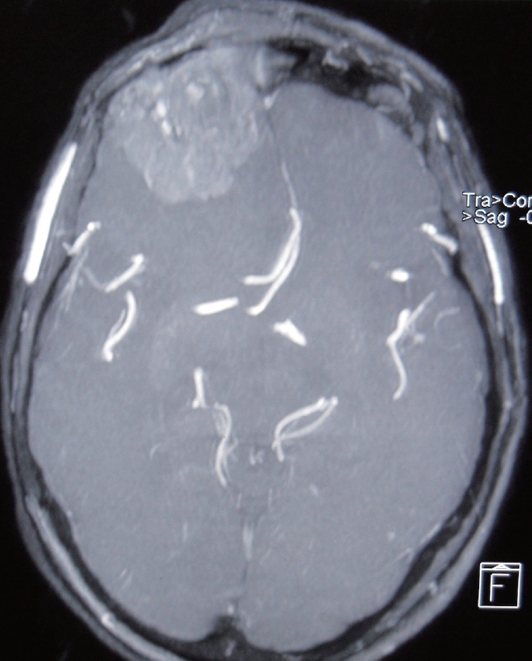
A 68-year-old female patient was admitted to the neurosurgical departmentãs inpatient clinic with chronic headache and rapidly progressive painful protruding mass on her head. Physical examination revealed a tender, 11û9 cm mass on the right frontal region of the skull. Her cranial nerve examination was normal. The skull survey showed large lytic changes in the frontal bone. Magnetic resonance imaging (MRI) of the head showed an extra-axial mass which was presumed to be a meningioma (Fig. 1). MRI angiography showed bilateral shift of the anterior cerebral arteries due to tumor mass. A frontal craniotomy was performed and the tumor was sub-totally removed. The mass was soft, highly vascular with a poorly defined border on the bone. The tumor mass was found to be attached to the dura with no intraparencymal spreading. The dura was intact at the time of operation.
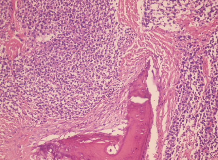
After removal, tumor tissue was fixed in 10% formalin solution, and dehydration was applied with graded alcohols and all of the specimen was embedded into paraffin. Hematoxylin-eosin stained sections of tumor revealed a monomorphous appearance of plasma cells with characteristic round-oval cells, eccentric nuclei, and abundant cytoplasm (Fig. 2). Immunohistochemical staining was conducted on the Ventana BenchMark XT (Roche Diagnostics, Basel, Switzerland) automated slide stainer. The tumor cells were positive for ö¤-chain, CD138 and CD38. However, they were negative for cytokeratin, CD45, CD20, ö£-chain, and CD3, revealing a pathological diagnosis of plasmacytoma.
The systemic evaluation required to make the diagnosis of solitary plasmacytoma included routine blood tests, serum calcium level and serum immunoglobulin levels, which were all normal. No Bence-Jones protein was detected in the urine. Bone marrow aspirate was unremarkable with normal morphological appearance and no bone marrow plasmacytosis. Computed tomography scan of the chest and abdomen showed no evidence of disease. A skeletal survey revealed no evidence of other bone lesions and bone scintigraphy showed that only the frontal bone was infiltrated by tumor.
Post-operative MRI showed eliminated midline shift and sub-totally removed tumor mass. The patient refused post-operative irradiation and systemic chemotherapy and was discharged. Four months later, she was re-admitted to our clinic with frontal swelling; MRI showed the re-growth of the previous mass. A new bone marrow biopsy showed no evidence of systemic disease and the skeletal survey was still negative for other lesions. The patient is alive and without any systemic dissemination of disease after 14 months.
DISCUSSION
Solitary plasmacytoma of bone consists about 5% of the malignant plasma cell tumors [8]. The spine, pelvis, and femur are the most common sites of involvement. However, the skull is a rare location for solitary plasmacytoma without signs of systemic myelomatosis [17]. Solitary craniocerebral plasmacytoma manifests in two forms : primary plasmacytomas arising from the skull and intracranial extramedullary plasmacytomas arising from the dura and the brain [10,15]. In order to diagnose solitary plasmacytoma, multiple myeloma must be ruld out, and the required systemic evaluation should include a skeletal survey, bone marrow aspirate, serum and urine protein electrophoresis and quantitative immunoglobulins.
Solitary plasmacytoma may occur at any age, but is mostly seen in patients in their fifties or sixties [7,16,19]. A complete blood count, complete metabolic profile and urinalysis with no abnormalities should be obtained. Clinical symptoms and signs are variable, non-specific and largely depend upon the location of tumor. The neurological abnormalities in patients with plasma cell neoplasms originating from the skull or dura are usually due to increased intracranial pressure and include headache, nausea, vomiting, along with visual disturbances [19]. Therefore, solitary craniocerebral plasmacytoma is often misdiagnosed pre-operatively. The differential diagnosis of the craniocerebral plasmacytoma includes meningioma, metastasis, lymphoma, osteochondroma, infectious meningitis and sarcoma [3]. Establishing the diagnosis of plasmacytoma requires a biopsy. Solitary plasmacytoma should be histologically distinguished from plasma cell granuloma. The plasma cell granuloma is characterized immunohistochemically by a polyclonal plasma-cell proliferation [5,20].
The etiology of plasmacytoma remains unclear. However, viral infections and trauma are thought to be involved in the etiology. Hepatitis C and Epstein-Barr viruses are possible viral agents influencing the development of plasmacytoma [11]. Our case had a history of head trauma about 10 years ago. A study by Pasch et al. [14] showed the possible role of previous trauma in plasmacytoma.
Surgical intervention is an excellent primary treatment option of solitary craniocerebral plasmacytoma. Arienta et al. [1] have reported that additional treatment is needed after the surgery. However (?), Jakubowski et al. [12] reported that radiotherapy is the most successful treatment after the surgery. Du Preez and Branca [10] reported that radiotherapy should be given only in cases with local recurrence. The cure rate has been suggested to be higher when radiotherapy (pre- or post-operative) was used together with surgical excision [3,10,17]. Our patient showed a recurrence 4 months after the initial diagnosis. We believe that the patientãs refusal to radiotherapy-chemotherapy treatment may be effective on early recurrence of this tumor
Solitary plasmacytoma of the bone tends to disseminate or progress to multiple myeloma over a period of 7-23 years [9,13,18]. Therefore, it should be kept in mind that solitary plasmacytoma may progress to multiple myeloma and strict follow-up of solitary plasmacytoma patients is of paramount importance.