Kim: Intraoperative Neurophysiology Monitoring for Spinal Dysraphism
Abstract
Spinal dysraphism often causes neurological impairment from direct involvement of lesions or from cord tethering. The conus medullaris and lumbosacral roots are most vulnerable. Surgical intervention such as untethering surgery is indicated to minimize or prevent further neurological deficits. Because untethering surgery itself imposes risk of neural injury, intraoperative neurophysiological monitoring (IONM) is indicated to help surgeons to be guided during surgery and to improve functional outcome. Monitoring of electromyography (EMG), motor evoked potential, and bulbocavernosus reflex (BCR) is essential modalities in IONM for untethering. Sensory evoked potential can be also employed to further interpretation. In specific, free-running EMG and triggered EMG is of most utility to identify lumbosacral roots within the field of surgery and filum terminale or non-functioning cord can be also confirmed by absence of responses at higher intensity of stimulation. The sacral nervous system should be vigilantly monitored as pathophysiology of tethered cord syndrome affects the sacral function most and earliest. BCR monitoring can be readily applicable for sacral monitoring and has been shown to be useful for prediction of postoperative sacral dysfunction. Further research is guaranteed because current IONM methodology in spinal dysraphism is still deficient of quantitative and objective evaluation and fails to directly measure the sacral autonomic nervous system.
Key Words: Monitoring, Intraoperative · Spinal dysraphism · Neural tube defect · Surgery.
INTRODUCTION
Spinal dysraphism is a complex entity of congenital anomalies of the spine and spinal cord. Tethered cord syndrome (TCS) is often induced from spinal dysraphism, as which anchors the spinal cord caudally and produces stretch injury [ 20]. Untethering surgery for the TCS from spinal dysraphism is indicated to prevent or partially reverse neurological impairment. However, surgical treatment involves risk of additional injury to the neural structures and application of the surgical treatment has always been weighed between the risk and benefit [ 10, 11]. Intra-operative neurophysiological monitoring (IONM) during untethering surgery plays an integral role to minimize the risk of neurological aggravation and maximize the efficacy of the surgery. Here in this review, principal knowledge about IONM for untethering surgery in spinal dysraphism is covered and clinical pearls from experience of the author will be shared.
BACKGROUND
Neuroanatomy related to spinal dysraphism
Spinal dysraphism refers to a spectrum of disorders and the detail is beyond the scope of the current review. Overall, spinal dysraphism results from impaired primary or secondary neurulation; although distinction may not be clear, myelomeningocele or lipomyelomeningocele, congenital dermal sinus, or limited dorsal myeloschisis occurs from defective primary neurulation and terminal myelocystocle, fatty filum terminale, or lumbosacral lipoma does from defective secondary neurulation [ 9]. The lower end of the cord including epiconus and conus medullaris comprises the motor neurons to the lower extremities, external urethral and anal sphincters, sensory tracts of the somatic nervous system, and the autonomic nucleus which gives off parasympathetic fibers to the urogenital and intestinal organs. On the contrary, sympathetic fibers innervating those organs originate from upper lumbar or thoracic levels and run through the sympathetic chain. The somatic nerve fibers form lumbosacral plexus and derive femoral, sciatic, obturator nerves to the lower limbs and the pudendal nerve to the pelvic organs. The autonomic fibers compose the superior and inferior hypogastric plexus and then contribute to vesical, rectal, or uterovaginal plexus via the sacral splanchnic nerves or pelvic splanchnic nerves. Thus, the course of the somatic nervous system and autonomic nervous system separate within the pelvic cavity whereas they adjoin at the cauda equina or conus level, except the sympathetic fibers. It is the very reason why monitoring of the external anal sphincter, innervated by the pudendal nerve, a somatic nervous system, can represent parts of the autonomic nervous system such as detrusor activity; most of neurological impairment in spinal dysraphism or TCS occurs at the level of conus medullaris or cauda equina.
Pathophysiology of TCS
There can be two mechanisms in the development of neurological impairment from TCS. First, structural anomalies often directly involve the conus or the cauda equina with corresponding neurological deficits. Alternatively, the spinal cord, anchored and stretched to the caudal end, undergoes neurological impairment by deranged oxidative metabolism and altered electrical properties of the neural membrane [ 19]. Direct structural damage can exhibit various neurological manifestation according to location and configuration of the lesion. Neurological impairment from cord traction preferentially develops sacral dysfunction, characteristic of TCS. Mechanical elongation of the cord induces decreased blood flow and neuronal membrane wrinkling, which leads to impaired oxidative metabolism and electrical activities. Due to the attachment to the dentate ligament, the more cranial level of the spinal cord is the less elongated; the sacral cord is more prone to traction injury than the lumbar or thoracic cord. Therefore, in TCS, sacral function, such as bladder, bowel, and sexual function, is compromised first and most, followed by motor and sensory functions of the lower extremities.
Intraoperative neurophysiological monitoring
The timing and technique of untethering surgery in spinal dysraphism has been subject to debate [ 11]. TCS can aggravate with growth of children while surgery itself involves risk of neural injury. Clinical utility and evidence of IONM for untethering will be explained later in detail. At any rate, untethering surgery in spinal dysraphism is intended to improve neurological outcome and thus IONM can be employed to evaluate neural function associated with TCS and to minimize neural injury during surgery. IONM should be individually considered because any lesion can never be the same. Protocols of IONM should be designed in correspondence to neurological status and risk in each patient. Nonetheless, because sacral nervous system is most vulnerable in TCS, sacral root level monitoring cannot be omitted. Lumbar root levels should be also monitored due to the concern of direct involvement of lesion or injury during untethering. From electrophysiological perspective, what IONM can directly monitor is the somatic nervous system, not the autonomic nervous system. In the somatic nervous system, afferent and efferent signals are entirely conveyed via electrical activities; efferent outputs in skeletal muscles including the external urethral and anal sphincters are also action potentials in sarcomere from excitation-contraction coupling. Meanwhile, in the autonomic nervous system, electrical signals are low in amplitude and slow in conduction and function of effector organs is not directly mediated by action potentials. For example, detrusor contraction is monitored by intravesical pressure rather than by action potentials [ 14]. Consequently, conventional IONM for untethering in spinal dysraphism focuses on the somatic nervous system of the lumbar and sacral levels. Monitoring the external anal sphincter motor and pudendal sensory function is the most often surrogate for the sacral nervous system. Moreover, the sympathetic nerves arising from the upper lumbar or thoracic spinal cord and traversing through the sympathetic chain and pelvic plexus cannot be represented in such ways.
METHODOLOGY
Electromyography (EMG) and motor evoked potential (MEP)
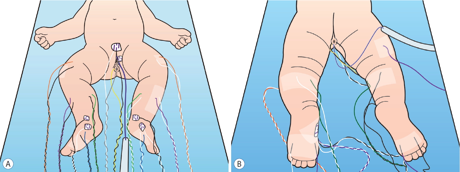
EMG and MEP are integral parts in IONM for untethering. It is because most of lumbosacral root levels can be monitored and identified by EMG and MEP. In this process, myotomal selection, or which muscles to sample, is a key question and can vary depending on expertise of electrophysiologists and neurological conditions of patients. Usually, vastus medialis, vastus lateralis, or rectus femoris is selected for L3, L4 level monitoring, tibialis anterior for L4, L5 monitoring, abductor hallucis for S1, S2 monitoring, and external anal sphincter (EAS) for S2 to S4 monitoring. The extensor hallucis longus can be added for monitoring of L5 level and biceps femoris or semitendinosus can be for S1, S2 levels. EMG and MEP are usually recorded by subdermal needle electrode inserted and secured in selected muscles. Surface disc electrode can be also used but it is not as effective to evaluate EMG signal, especially to discern neurotonic discharges related to neural injury from phasic discharges [ 12] ( Fig. 1). EMG monitoring can be classified into free-running EMG and triggered EMG. Free-running EMG is useful to find out whether current surgical manipulation is irritating neural tissue in vicinity. Phasic discharges are frequently observed as an irregular, transient electrical signals and indicate irritation without injury. Neurotonic discharges are high-frequency, regular, and persistent signals and signifies neural injury. Triggered EMG is employed for mapping of nerve root or rootlets. By applying electrical stimulation with probe stimulator and recording EMG signals from any muscles, surgeons can identify presence of neural tissue at the site of stimulation and corresponding root level. Bipolar concentric probe stimulator is recommended for stimulation in triggered EMG because it induces less current spread to nearby tissue thus yielding better specificity. Stimulation intensity should be considered and optimized for sensitivity and specificity and protocols can vary per surgeons and settings. Usually, pulse duration can be set between 75 and 500 µsec and current intensity can be modified for each stimulation from 0.1 to 10 mA. Direct neural stimulation produces EMG response at 0.5 to 2 mA while absence of response at higher intensity (up to 10 mA) excludes presence of neural tissue at the site of stimulation.
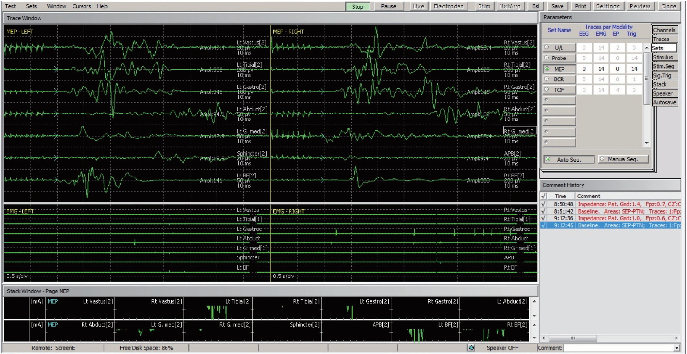
MEP is also monitored from the same electrodes installed for EMG monitoring. Stimulation is delivered by transcranial electrical stimulation via subdermal needle electrodes in the scalp. Arrangement of electrodes (also called as “montage”) is recommended at C3 and C4 in the scalp for monitoring upper or lower extremity MEP; Cz-Fz montage or C1-C2 montage can be also considered for lower extremity MEP stimulation but C3-C4 montage is more efficacious to yield lower extremity MEP than C1-C2 montage [ 6] and Cz-Fz electrodes are usually spared for sensory evoked potentials. Stimulation intensity should be maintained at the supra-maximal level. Decrease of MEP amplitude together with prolonged neurotonic discharge in free-running EMG anticipates corresponding neurological deficits post-operatively ( Fig. 2).
Filum terminale and cord stimulation
Identification of the filum terminale or non-functioning cord tissue is as important as identification of nerves in order to achieve safe untethering by sectioning or debulking of them. It should be confirmed that target tissue does not contain neural elements before removal. Inadvertent removal of neural tissues will result in post-operative neurological impairment. Triggered EMG at higher intensity (up to 10 mA) should not generate EMG signals to guarantee absence of the nerves. As for the filum terminale, 1 : 100 rule has been suggested; stimulation threshold of the filum terminale is 100 times higher than that of the roots or rootlets [ 13, 18]. Based on the experience of the author and colleagues, 10 mA stimulation is enough for that purpose in most of cases.
Sensory evoked potential
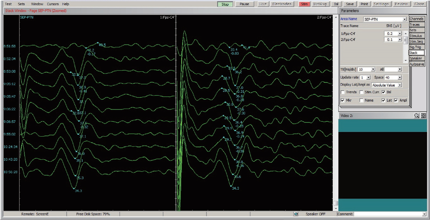
Clinical utility of sensory evoked potential (SEP) is limited compared to that of EMG or MEP in untethering surgery. It is because conventional SEP from posterior tibial nerve of the lower extremity does not cover sufficient root levels of concern and the change of SEP cannot be immediately recognized. Posterior tibial nerve SEP can be omitted when number of channels is limited in IONM equipment ( Fig. 3). However, pudendal SEP can be useful for interpretation of sacral monitoring [ 4]. Sacral function is effectively monitored by bulbocavernosus reflex (BCR) and EAS MEP. BCR covers both the motor and sensory nervous pathway in the sacral root level and EAS MEP represents the motor pathways to the sacral root level. Additionally, pudendal SEP evaluates the sensory pathways from the sacral roots. In cases when BCR is compromised, further tests of EAS MEP or pudendal SEP can differentiate which pathway, motor versus sensory, is being impaired. Or, if BCR or EAS MEP is not reliable or obtainable from baseline, pudendal SEP can be employed for sacral monitoring.
BCR
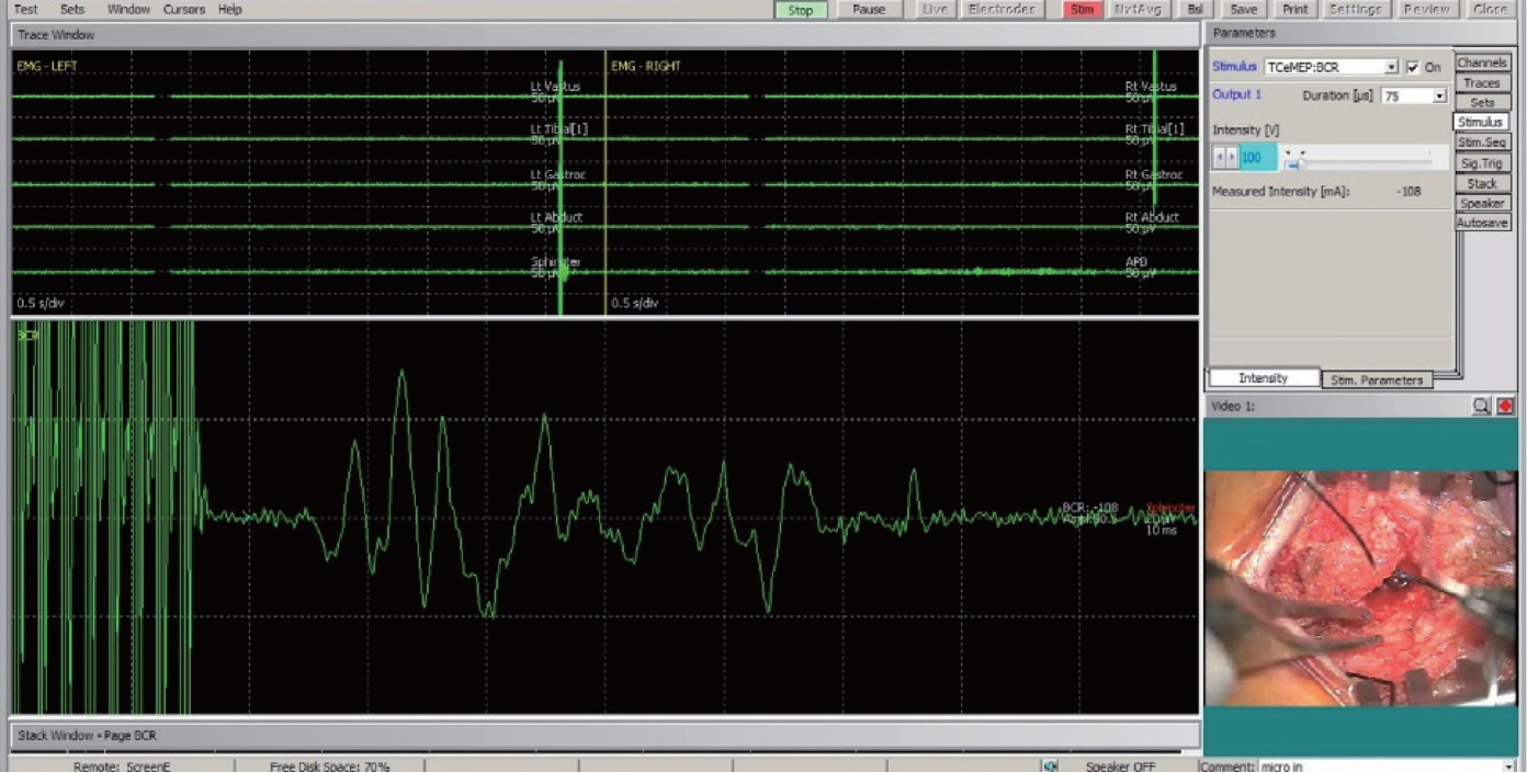
BCR is of the greatest utility for the monitoring of sacral function in untethering surgery [ 5]. It is performed by electrically stimulating pudendal afferent fibers at the clitoris or penis using surface electrode and recording triggered EMG in EAS using subdermal needle electrode [ 3]. The reflex arc of BCR covers both sensory and motor fibers in the conus and sacral roots. Therefore, change of BCR is highly specific to predict post-operative sacral function including voiding; if BCR is preserved, post-operative voiding is maintained in about 90% at 6 months after surgery [ 2] ( Fig. 4).
Pudendal sensory nerve action potential
Routinely, the above IONM techniques (EMG, MEP, SEP, and BCR) are clinically sufficient for untethering. Additionally, pudendal sensory nerve action potential was suggested to monitor to identify pudendal sensory fibers. Pudendal sensory nerve action potential is recorded by hand-held bipolar tip electrode applied to target tissue by surgeon and stimulated at the penis or clitoris in the same way as in BCR [ 7]. It requires extra effort from surgeon and takes time to obtain averaged response. Moreover, sensory pathways can be effectively monitored from BCR and can be identified as well by recording triggered EMG from EAS delivered through reflex arc. Thus, there is not much clinical necessity for pudendal sensory fibers to be identified in isolation.
CLINICAL SIGNIFICANCE
With regard to clinical evidence of IONM for untethering in spinal dysraphism, randomized controlled trials that compared application of IONM are not available as many other conditions. However, whether application or IONM for untethering is advantageous is not a subject to debate at present. Concerns had remained whether the surgery should be performed ahead of growth or after the occurrence of neurological deficits because of risk of further neurological impairment from surgery. However, along with advance of IONM, surgical intervention in spinal dysraphism is now generally accepted to be indicated early before neurological compromise. Early surgery is expected to lead to superior functional outcome in long-term [ 11]. Although other confounding factors are not controlled, evidences exist that showed untethering with use of IONM provided better clinical outcome in terms of pain, sensorimotor function, and sphincter function [ 13]. Of interest, one report is available that compared surgical outcome of three surgeons in a single center [ 1]. In the report, only the surgeon who employed IONM in the fewest cases had observed postoperative neurological deficits. Especially monitoring EMG and MEP from external anal or urethral sphincters and lower extremities were shown to decrease neurological deterioration after untethering surgery [ 18]. However, posterior tibial SEP has not been shown to prevent neurological deficits [ 8, 15]. However, it is still admitted that EMG, MEP, and SEP may fail to effectively monitor autonomic functions including detrusor activity [ 15].
CLINICAL PEARLS
Based on literature review and clinical experience, personal recommendations can be made for IONM in spinal dysraphism surgery as below. EMG and MEP monitoring is imperative and it should cover as much root levels as required. In our hospital, EMG and MEP monitoring spans from mid lumbar level to sacral level by including vastus medialis, tibialis anterior, gastrocnemisus, abductor hallucis, and EAS ( Fig. 1). Electrodes are installed for SEP monitoring, either tibial or pudendal, at Fz- Cz montage on the scalp. At baseline, tibial or pudendal SEPs are evaluated but need not be continuously monitored because of less clinical utility. Only when neurological impairment is suspected from EMG, MEP, or BCR, pudendal or tibial SEP is checked to confirm or further localize a corresponding lesion. BCR is always emphasized to evaluate sacral function. Baseline test of BCR can reveal current neurological status of patients and change of BCR during surgery highly correlates with progression of neurological impairment at the sacral root level. Before sectioning or removal of lesion, triggered EMG is performed at high intensity (~10 mA) to confirm the absence of functional neural tissue. Interpretation of IONM signals in untethering surgery may be further modified from general IONM protocols. Free-running EMG observes tonic or phasic EMG signals more often than other surgeries. Appearance of EMG signals did not highly correlate with post-operative neurological deficits. Rather, it could be regarded as signifying that corresponding rootlets may reside near current site of manipulation. Change of MEP amplitude or morphology is more specific in untethering surgery compared to other spinal cord surgeries. It can be explained by the fact that most of muscles sampled are innervated by two or more root levels and injury to a single root level may not produce disappearance of MEP which is accepted as clinically significant change in other spine surgeries [ 17]. Thus, in surgery for spinal dysraphism, neurophysiologists should be more cautious about change of MEP even without complete loss.
FURTHER RESEARCH
Current techniques of IONM for untethering in spinal dysraphism is effective to guide surgeons and to improve clinical outcome. However, there is still a room for research and development. First of all, one of integral functions of sacral cord is autonomic regulation of the bladder, bowel, and genital organs. However, electrophysiological tests are not efficient for monitoring of the autonomic nervous system because the effector organs do not function electrically as in the somatic nervous system. Detrusor function can be monitored using intraoperative cystometry which measures intravesical pressure in real-time [ 14]. However, the response is delayed and less quantitative. There has been a recent research which attempted to electrophsyiologically measure sexual function during urological surgery [ 16]. Basic research is mandatory to conceive a novel methodology for monitoring of the autonomic nervous system. Second, interpretation of IONM responses are still much dependent on electrophysiologists’ discretion. For example, whether a certain spontaneous EMG signal is pathologic or benign is determined subjectively in the context of lesion extent or surgical process. Significant changes of MEP or SEP are also decided differently by institute, surgeon, or surgery. More quantitative and objective monitoring would be possible with better understanding of neurophysiology and development of related biomedical engineering.
Additionally, although we may be inclined to believe that neuroanatomy or neurophysiology of the spinal cord and roots are unveiled enough for clinical purpose, I as an electrophysiologist feel much of the knowledge is still unexplored. Manipulative investigation on stimulation and response of the nervous system is guaranteed for future study.
CONCLUSION
IONM for surgery of spinal dysraphism is indicated to guide surgeons during surgery and to minimize neurological deficits. The sacral nervous system is most vulnerable to neurological impairment from TCS and should be monitored with caution. Free-running EMG and triggered EMG is useful to identify lumbosacral roots within the field of surgery. Filum terminale or non-functioning cord can be confirmed by absence of responses at higher intensity of stimulation. BCR monitoring is predictive of post-operative sacral dysfunction. SEP can be also employed to further interpretation.
Acknowledgements
I sincerely and deeply appreciate Prof. Kyu-Chang Wang of his devotion and guidance to intraoperative neurophysiological monitoring in pediatric neurosurgery. I entered into the field by his consideration and could develop it with lessons from him. Intraoperative monitoring of spinal dysraphism surgery was the very topic where I started. I am certainly confident that our intraoperative monitoring is at the highest level, comparable to any renowned institute world-wide, solely thanks to him.
Fig. 1.
Muscles selected to monitor electromyography and motor evoked potential, in front (A) and rear (B) view. Vastus medialis, tibialis anterior, abductor hallucis, gastrocnemius, and external anal sphincter was installed with subdermal needle electrodes. 
Fig. 2.
Motor evoked potentials and free-running electromyography monitored during untethering surgery. 
Fig. 3.
Sensory evoked potentials from posterior tibial nerve monitored during untethering surgery. 
Fig. 4.
Bulbocavernosus reflex monitored during untethering surgery. 
References
1. Albright AL, Pollack IF, Adelson PD, Solot JJ : Outcome data and analysis in pediatric neurosurgery. Neurosurgery 45 : 101-106, 1999   2. Cha S, Wang KC, Park K, Shin HI, Lee JY, Chong S, et al : Predictive value of intraoperative bulbocavernosus reflex during untethering surgery for post-operative voiding function. Clin Neurophysiol 129 : 2594-2601, 2018   3. Deletis V, Vodusek DB : Intraoperative recording of the bulbocavernosus reflex. Neurosurgery 40 : 88-93; discussion 92-93, 1997   4. Deletis V, Vodusek DB, Abbott R, Epstein FJ, Turndorf H : Intraoperative monitoring of the dorsal sacral roots: minimizing the risk of iatrogenic micturition disorders. Neurosurgery 30 : 72-75, 1992   5. Hwang H, Wang KC, Bang MS, Shin HI, Kim SK, Phi JH, et al : Optimal stimulation parameters for intraoperative bulbocavernosus reflex in infants. J Neurosurg Pediatr 20 : 464-470, 2017   6. Journée HL, Polak HE, de Kleuver M : Influence of electrode impedance on threshold voltage for transcranial electrical stimulation in motor evoked potential monitoring. Med Biol Eng Comput 42 : 557-561, 2004    7. Kothbauer KF, Novak K : Intraoperative monitoring for tethered cord surgery: an update. Neurosurg Focus 16 : E8, 2004  8. Krassioukov AV, Sarjeant R, Arkia H, Fehlings MG : Multimodality intraoperative monitoring during complex lumbosacral procedures: indications, techniques, and long-term follow-up review of 61 consecutive cases. J Neurosurg Spine 1 : 243-253, 2004   9. McLone DG : Occult dysraphism and the tethered spinal cord lipomas. Edited by Choix M, Di Rocco C, Hockley A : In: Pediatric Neurosurgery. Philadelphia : Churchill Livingstone, 1999, pp61-78
10. Pang D, Zovickian J, Oviedo A : Long-term outcome of total and near-total resection of spinal cord lipomas and radical reconstruction of the neural placode: part I-surgical technique. Neurosurgery 65 : 511-528, 2009    11. Pang D, Zovickian J, Oviedo A : Long-term outcome of total and near-total resection of spinal cord lipomas and radical reconstruction of the neural placode, part II: outcome analysis and preoperative profiling. Neurosurgery 66 : 253-272, 2010    12. Prell J, Rampp S, Romstöck J, Fahlbusch R, Strauss C : Train time as a quantitative electromyographic parameter for facial nerve function in patients undergoing surgery for vestibular schwannoma. J Neurosurg 106 : 826-832, 2007   13. Quiñones-Hinojosa A, Gadkary CA, Gulati M, Von Koch CS, Lyon R, Weinstein PR, et al : Neurophysiological monitoring for safe surgical tethered cord syndrome release in adults. Surg Neurol 62 : 127-133, 2004   14. Ryken T, Menezes A : Intraoperative electrical and manometric monitoring in lumbosacral surgery. Edited by Loftus CM, Traynelis VC : In: Intraoperative Monitoring Techniques in Neurosurgery. New York : McGraw-Hill, 1994, pp257-268
15. Shinomiya K, Fuchioka M, Matsuoka T, Okamoto A, Yoshida H, Mutoh N, et al : Intraoperative monitoring for tethered spinal cord syndrome. Spine (Phila Pa 1976) 16 : 1290-1294, 1991   16. Song WH, Park JH, Tae BS, Kim SM, Hur M, Seo JH, et al : Establishment of novel intraoperative monitoring and mapping method for the cavernous nerve during robot-assisted radical prostatectomy: results of the phase I/II, first-in-human, feasibility study. Eur Urol 78 : 221-228, 2020  17. Tanaka Y, Kawaguchi M, Noguchi Y, Yoshitani K, Kawamata M, Masui K, et al : Systematic review of motor evoked potentials monitoring during thoracic and thoracoabdominal aortic aneurysm open repair surgery: a diagnostic meta-analysis. J Anesth 30 : 1037-1050, 2016    18. Von Koch CS, Quinones-Hinojosa A, Gulati M, Lyon R, Peacock WJ, Yingling CD : Clinical outcome in children undergoing tethered cord release utilizing intraoperative neurophysiological monitoring. Pediatr Neurosurg 37 : 81-86, 2002   19. Yamada S, Won DJ, Yamada SM : Pathophysiology of tethered cord syndrome: correlation with symptomatology. Neurosurg Focus 16 : E6, 2004  20. Yamada S, Zinke DE, Sanders D : Pathophysiology of “tethered cord syndrome”. J Neurosurg 54 : 494-503, 1981   
|
|


