| Home | E-Submission | Sitemap | Editorial Office |  
top_img
Ki, Lee, Hur, and Lee: Post-Traumatic Arteriovenous Fistula of the Scalp

Abstract

Arteriovenous fistula of the scalp is relatively rare disease. We report a traumatic arteriovenous fistula of the scalp treated with complete surgical excision and review the literature with regard to etiology, pathogenesis, and management of these unusual lesions.

INTRODUCTION

Traumatic arteriovenous fistulas (AVFs) of the scalp are rarely encountered by the neurosurgeon. AVF of the scalp is the direct connection between the arterial feeding vessels of the scalp and the draining veins without intervening capillary beds. The draining veins or feeding arteries are often grossly dilated, creating a cosmetic deformity. The symptoms are pulsatile mass, bruit, tinnitus, and headache8). The treatment options include surgical excision1,24,67,811,1214,1516,1722), ligation of feeders, transarterial and transvenous embolization5,910,1323), and injection of sclerosing agent21). We experienced a case of traumatic AVF of scalp, successfully treated by surgical excision.

CASE REPORT

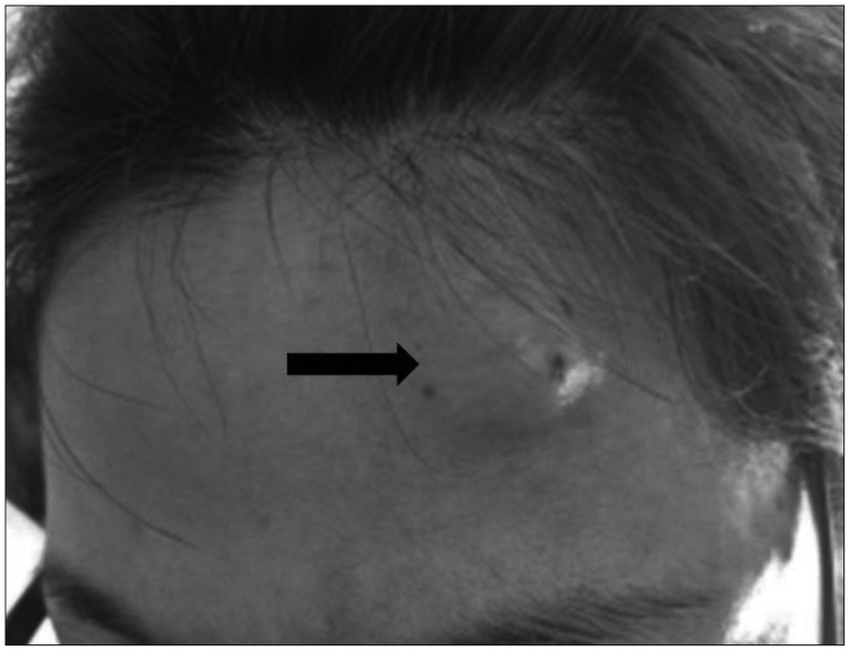
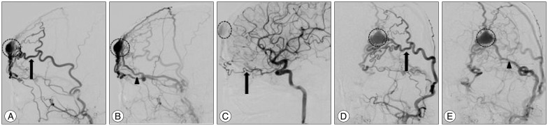
A 42-year-old woman bumped her forehead against the TV about 30 years ago. She noticed the swelling at her left forehead after the trauma; thereafter, a small lesion persisted without any symptoms or size alteration. From 4-5 years ago, "the mass" has gradually increased in size, and showed pulsation. She could hear bruit from the mass when she lay down; the patient also complained about frequent headaches. Physical examination revealed 25×20 mm-sized pulsatile and a round mass on the left forehead (Fig. 1). There was a continuous bruit on auscultation. The pulsation of the mass disappeared with the compression on the proximal portion of the superficial temporal artery (STA). The presumptive diagnosis of AVF was confirmed by selective internal and external carotid digital subtraction angiography (Fig. 2). The main arterial supply was from the left STA, and other feeders were the left deep temporal artery and the ethmoid branch of the left ophthalmic artery. Drainage was seen preferentially into the superficial temporal vein, as well as into the left frontal cortical veins and left superior ophthalmic vein.
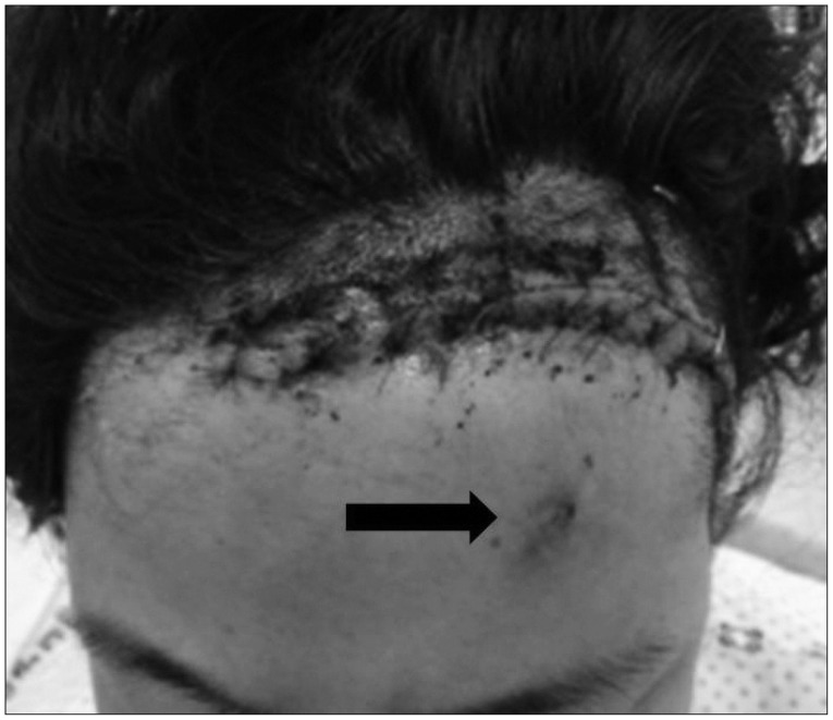
The operation was performed under general anesthesia. A curvilinear 10 cm skin incision was made on the inner part of hairline because of a cosmetic problem. The right STA and AVF were gently dissected from the adjacent subcutaneous tissue. The largest feeder was from the STA and was controlled with a hemoclip. Other vessels were all coagulated and cut. After operation, all the symptoms and signs disappeared, and no other complications developed (Fig. 3). The pathologic report confirmed consistent AVF.

DISCUSSION

The AVF of the scalp has been previously named cirsoid aneurysm, rasemose aneurysm, plexiform angioma, pulsating anigoma, aneurysmal varix, arteriovenous aneurysm, and arteriovenous malformation8,17). The location of this lesion is roughly evenly distributed among the frontal, temporal, parietal, and occipital regions6). The etiology of the AVF of the scalp is still controversial, however, it may be either of congenital or traumatic origin. Among 64 patients with the AVF of the scalp reported in previous research1,23,45,67,89,1011,1213,1415,1617,1821,2223), 36 patients had the traumatic origin. Traumatic origin splits into incidental injury (28 patients) and iatrogenic injury (8 patients). Iatrogenic injury included hair transplantation3,45), craniotomy1,16), and temporomandibular joint arthroscopy5). Theories about pathogenesis of the congenital AVF include persistence of primitive AVFs, as a sequel to vascular hamartomas, and formation at a site of arteriovenous crossing8). On the other hand, there are two theories about pathogenesis of the traumatic AVFs. One is a disruption of the arterial wall and its vasa vasorum with endothelial proliferation to adjacent veins2). The other is simultaneous lacerations of the artery and the accompanying vein result in a single fistula20).
Management of scalp AVF is difficult because of its high shunt flow, complex vascular anatomy, and associated cosmetic problems. The indication of treatment includes cosmetic relief of the pulsatile mass, prevention of hemorrhage, as well as other symptoms such as headache and tinnitus8). In the majority of cases1,24,67,811,1214,1516,1722), radical surgical excision has been the most common treatment method. Complete excision of the fistula with ligation of all feeding vessels is necessary, as there is a high propensity of these lesions to recur. Incomplete treatment can also cause scalp necrosis and bleeding. Additionally, primary scalp vascular malformation can be excised entirely without significant blood loss and scalp necrosis. However, excision of secondary scalp venous dilatation without treatment of the intracranial component can be life-threatening19). Other therapeutic methods, such as ligation of feeding vessels, transarterial and transvenous embolization5,910,1323), and injection of sclerosing agent21) have been used to treat AVF. In the reported case, we choose the surgical method because of the cosmetic issue and the probability of recurrence with other methods. We applied a hemoclip on the largest feeder (STA), and sacrificed the other vessels with coagulation. No complications were observed, and the patient was satisfied with the outcome.

CONCLUSION

Traumatic AVF of the scalp must be considered if the patient had a trauma and has a palpable thrill and/or pulsatile mass lesion, disappeared after proximal STA compression. There are many different methods to treat these lesions. In the present study, surgical excision under general anesthesia was chosen.

References

1. Amlashi SF, Riffaud L, Morandi X : Arteriovenous fistula of the superficial temporal artery : an exceptional complication of the pterional approach. J Neurol Neurosurg Psychiatry 2004, 75 : 1077-1078,
crossref
2. Badejo L, Rockwood P : Traumatic arteriovenous fistula of the scalp. Case report. J Neurosurg 1987, 66 : 773-774,
crossref pmid
3. Bernstein J, Podnos S, Leavitt M : Arteriovenous fistula following hair transplantation. Dermatol Surg 2011, 37 : 873-875,
crossref pmid
4. Champeaux C, Jecko V, Penchet G, Chibbaro S : Scalp arteriovenous fistula following hair transplantation. Clin Neuroradiol 2014, 24 : 285-288,
crossref pmid
5. Dabus G, Pizzolato R, Lin E, Kreusch A, Linfante I : Endovascular treatment for traumatic scalp arteriovenous fistulas : results with Onyx embolization. J Neurointerv Surg 2014, 6 : 405-408,
crossref pmid
6. El Shazly AA, Saoud KM : Results of surgical excision of cirsoid aneurysm of the scalp without preoperative interventions. Asian J Neurosurg 2012, 7 : 191-196,
crossref pmid pmc
7. Elkin DC : Cirsoid aneurism of the scalp : with the report of an advanced case. Ann Surg 1924, 80 : 332-340,
crossref pmid pmc
8. Fisher-Jeffes ND, Domingo Z, Madden M, de Villiers JC : Arteriovenous malformations of the scalp. Neurosurgery 1995, 36 : 656-660, discussion 660
crossref pmid
9. Heilman CB, Kwan ES, Klucznik RP, Cohen AR : Elimination of a cirsoid aneurysm of the scalp by direct percutaneous embolization with thrombogenic coils. Case report. J Neurosurg 1990, 73 : 296-300,
crossref pmid
10. Hendrix LE, Meyer GA, Erickson SJ : Cirsoid aneurysm treatment by percutaneous injection of sodium tetradecyl sulfate. Surg Neurol 1996, 46 : 557-560, discussion 560-561
crossref pmid
11. Hyshaw C, DiTullio M, Renaudin J : Superficial temporal arteriovenous fistula. Surg Neurol 1979, 12 : 46-48,
pmid
12. Johns DR, Swann KW, Heros RC : Spontaneous arteriovenous fistula of the superficial temporal artery. Surg Neurol 1987, 27 : 273-276,
crossref pmid
13. Kumar A, Ahuja CK, Khandelwal N, Bakshi JB : Cirsoid aneurysm of the right pre-auricular region : an unusual cause of tinnitus managed by endovascular glue embolisation. J Laryngol Otol 2012, 126 : 923-927,
crossref pmid
14. Li F, Zhu S, Liu Y, Chen Y, Chi L, Chen G, et al : Traumatic arteriovenous fistula of the superficial temporal artery. J Clin Neurosci 2007, 14 : 595-600,
crossref pmid
15. Morandi X, Godey B, Riffaud L, Brassier G : Nontraumatic arteriovenous fistula of the superficial temporal artery. Otolaryngol Head Neck Surg 2001, 124 : 588-589,
crossref pmid
16. Morioka T, Nishio S, Hikita T : Traumatic arteriovenous fistulae of the scalp at the area of previous craniotomy. Surg Neurol 1988, 30 : 406-407,
crossref pmid
17. Muthukumar N, Rajagopal V, Manoharan AV, Durairaj N : Surgical management of cirsoid aneurysms. Acta Neurochir (Wien) 2002, 144 : 349-356,
crossref pmid
18. Saade C, Wilkinson M, Parker G, Dubenec S, Brennan P : Multidetector computed tomography in the evaluation of cirsoid aneurysm of the scalp--a manifestation of trauma. Clin Imaging 2013, 37 : 558-560,
crossref pmid
19. Shenoy SN, Raja A : Scalp arteriovenous malformations. Neurol India 2004, 52 : 478-481,
pmid
20. Stasik JF, Engelcke BR, Zwemer F, Tomaro AJ : False aneurysm of the external maxillary artery : report of case. J Oral Surg Anesth Hosp Dent Serv 1964, 22 : 441-445,
pmid
21. Whiteside OJ, Monksfield P, Steventon NB, Byrne J, Burton MJ : Endovascular embolization of a traumatic arteriovenous fistula of the superficial temporal artery. J Laryngol Otol 2005, 119 : 322-324,
crossref pmid
22. Yi WJ, Cho JH, Lee HJ, Lee HG, Ryu KY, Kang DG, et al : Surgical treatment of a post-traumatic arteriovenous fistula of the superficial temporal artery and vein. J Korean Neurotraumatol Soc 2007, 3 : 59-61,
crossref
23. Youn SW, Lee NJ, Suh SI, Kang SH : Direct-puncture embolization of scalp arteriovenous fistulae. Neurol Med Chir (Tokyo) 2012, 52 : 525-528,
crossref pmid
Fig. 1

The clinical photo showing 25×20 mm-sized round mass on left forehead (arrow).

jkns-58-298-g001.jpg
Fig. 2

Digital subtraction angiographic images depicting the arteriovenous fistula : selective left external carotid artery from which the left superficial temporal artery comes (arrow) and drains itself mainly through the fistula (dashed circle) into multiple large superficial temporal veins (arrowhead). A and B : Lateral views. D and E : Front views. C : Lateral view of the left internal carotid artery showing feeding artery from branch of left opthalmic artery (arrow).

jkns-58-298-g002.jpg
Fig. 3

The post-operative photo showing the totally removed mass (arrow) and suture line.

jkns-58-298-g003.jpg
Editorial Office
1F, 18, Heolleung-ro 569-gil, Gangnam-gu, Seoul, Republic of Korea
TEL: +82-2-525-7552   FAX: +82-2-525-7554   E-mail: office@jkns.or.kr
About |  Browse Articles |  Current Issue |  For Authors and Reviewers
Copyright © Korean Neurosurgical Society.                 Developed in M2PI
Close layer