| Home | E-Submission | Sitemap | Editorial Office |  
top_img
Antar and Turk: Additional Surgical Method Aimed to Increase Distractive Force during Occipitocervical Stabilization : Technical Note

Abstract

Objective

Craniovertebral junctional anomalies constitute a technical challenge. Surgical opening of atlantoaxial joint region is a complex procedure especially in patients with nuchal deformity like basilar invagination. This region has actually very complicated anatomical and functional characteristics, including multiple joints providing extension, flexion, and wide rotation. In fact, it is also a bottleneck region where bones, neural structures, and blood vessels are located. Stabilization surgery regarding this region should consider the fact that the area exposes excessive and life-long stress due to complex movements and human posture. Therefore, all options should be considered for surgical stabilization, and they could be interchanged during the surgery, if required.

Methods

A 53-year-old male patient applied to outpatients’ clinic with complaints of head and neck pain persisting for a long time. Physical examination was normal except increased deep tendon reflexes. The patient was on long-term corticosteroid due to an allergic disease. Magnetic resonance imaging and computed tomography findings indicated basilar invagination and atlantoaxial dislocation. The patient underwent C0-C3-C4 (lateral mass) and additional C0-C2 (translaminar) stabilization surgery.

Results

In routine practice, the sites where rods are bound to occipital plates were placed as paramedian. Instead, we inserted lateral mass screw to the sites where occipital screws were inserted on the occipital plate, thereby creating a site where extra rod could be bound. When C2 translaminar screw is inserted, screw caps remain on the median plane, which makes them difficult to bind to contralateral system. These bind directly to occipital plate without any connection from this region to the contralateral system. Advantages of this technique include easy insertion of C2 translaminar screws, presence of increased screw sizes, and exclusion of pullout forces onto the screw from neck movements. Another advantage of the technique is the median placement of the rod; i.e., thick part of the occipital bone is in alignment with axial loading.

Conclusion

We believe that this technique, which could be easily performed as adjuvant to classical stabilization surgery with no need for special screw and rod, may improve distraction force in patients with low bone density.

INTRODUCTION

Craniovertebral junctional anomalies constitute a technical challenge. Surgical opening of atlantoaxial joint region is a complex procedure especially in patients with nuchal deformity like basilar invagination9). This region has actually very complicated anatomical and functional characteristics, including multiple joints providing extension, flexion, and wide rotation. In fact, it is also a bottleneck region where bones, neural structures, and blood vessels are located17).
Stabilization surgery regarding this region should consider the fact that the area exposes excessive and life-long stress due to complex movements and human posture8). Therefore, all options should be considered for surgical stabilization, and they could be interchanged during the surgery, if required. Surgical treatment of this region is composed of decompression and stabilization. Bone quality of the surgical candidate is very critical for stabilization.
We aimed to report on a surgical technique performed adjuvant to the standard occipitocervical stabilization surgery to improve distractive force in a patient with low bone quality.

CASE REPORT

Case

A 53-year-old male patient applied to outpatients’ clinic with complaints of head and neck pain persisting for a long time. Physical examination was normal except increased deep tendon reflexes. The patient was on long-term corticosteroid due to an allergic disease. Magnetic resonance imaging (MRI) and computed tomography (CT) findings indicated basilar invagination and atlantoaxial dislocation (Fig. 1). Bone densitometry showed a total T-score of -2.3 in lumbar region.

Surgery

The patient underwent C0-C3-C4 (lateral mass) and additional C0-C2 (translaminar) stabilization surgery.

Technique

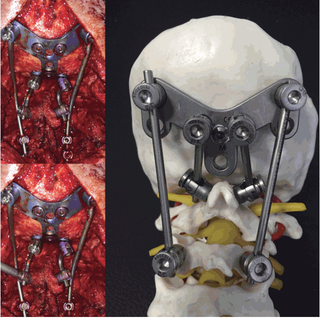
The technique can be described as the binding to the occipital plate by extra rod without a need for additional kit between C2 translaminar and C0, that was performed as adjuvant to C0-C4 lateral mass and pedicle screw stabilization surgery. In routine practice, the sites where rods are bound to occipital plates were placed as paramedian. Instead, we inserted polyaxial screw to the sites where occipital screws were inserted on the occipital plate, thereby creating a site where extra rod could be bound. When C2 translaminar screw is inserted, screw caps remain on the median plane, which makes them difficult to bind to contralateral system. These bind directly to occipital plate without any connection from this region to the contralateral system.
Advantages of this technique include easy insertion of C2 translaminar screws, presence of increased screw sizes, and exclusion of pullout forces onto the screw from neck movements. Another advantage of the technique is the median placement of the rod; i.e., thick part of the occipital bone is in alignment with axial loading.
This technique could allow for more powerful axial loading by independent insertion from the existing system in such surgeries where C2 lamina is preserved. Besides, it does not require any additional kit or design since screws used in occipital region are already included in the current kit. In addition, the rod may be bound to desired region since screw holes on the occipital plates are used (Fig. 2).

DISCUSSION

Craniovertebral junction is a complex structure due to presence of bones, neural tissues, and arteries. Mobilization of this region is provided by involvement of multiple joints. Therefore, surgical success rate depends on a successful stabilization, where many surgical techniques have been tried so far.
We hereby report an easy-to-perform surgical technique that may be done as adjuvant to previously defined surgical techniques.
Stabilization surgery of this region can be divided into two, as anterior and posterior stabilization17). In fact, higher number of surgical technique was described for the latter. Interlaminar grafting and wiring methods in posterior atlantoaxial fixation was first described by Gallie, which was then modified by Brooks and Jenkins1) and others2,6). Failure of these techniques was highly probable due to relaxation of wires and melting the bony graft inserted. These led to development of plate and rod systems. Goel and Laher were the first to describe the stabilization of C2 and C1 lateral mass with anchor screw, which required cutting of C2 root to stabilize C1 and C2 with plate7). In addition, Harms and Melcher5) in their study of 2001 used polyaxial screw instead of anchor screw on C2, and fixed it with the rod. The technique was further modified by replacement with translaminar screw by Wright in 2004, which was called as Goel-Harms-Wright method14). Except this method, screw stabilization of C1 lateral mass include also Tan method18) and Notch method11). Cutting of C2 nerve root may be a shared feature of all these techniques. C2 pedicle screw ensures a relatively rigid stabilization, which constitutes the first option for this region. Major disadvantage of this screw is that pedicle diameter could not have a small size and it is anteriorly related to carotid artery, which makes it prone to injury by screw’s tip17). Stabilization with transarticular screw was first developed by Mayers and Seeman in 1987. This method provides very rigid fixation with high fusion rates12,13).
Limitations of this technique include high riding vertebral artery, pedicle thickness, and insertion angle of screw3). C2 translaminar screw was first described by Wright in 200419). It might be used in patients where stabilization with C2 pedicle screw was troublesome (in case of high riding vertebral artery or small pedicle diameter). Biomechanistic studies showed it had comparable stability to pedicle screw10). Advantages of translaminar screw are ease of applicability, no need for fluoroscopy, no relation with vertebral artery, and similar amount of stabilization as pedicle screw. The only disadvantage is the difficulty of connection to other systems. Occipitocervical stabilization was first performed in 1980s with Ransford loop as the standard method15,16). This method was performed by wire-binding of shaped rod to the occipital bone and cervical laminae. After 20 years of usage, the Olerud cervical fixation system was introduced in 1997. Short segment fixation with C0-C2 translaminar screw or pedicle screw provides a very strong stabilization in patients stabilized under C2. Similarly, their midline or bicortical placements were very strong in the relation of occipital plate to the occipital bone4).
In addition, there are hybrid techniques in the literature where translaminar screw and various connections are simultaneously used3,14,20). In our case, we tried to strengthen the stabilization by adding two rods to the midline adjuvant to stabilization of the occipital bone with classical pedicle and lateral mass. Postoperative CT and MRI images showed narrowed atlantodental interval with relief of compression on the spinal nerve (Fig. 3). This method may be added to the occipitocervical stabilization by 2 extra rods without a need for any additional material. Moreover, rod may be connected to the desired site of the occipital plate, and difficulties regarding rod connection could be overcome.

CONCLUSION

We believe that this technique, which could be easily performed as adjuvant to classical stabilization surgery with no need for special screw and rod, may improve distraction force in patients with low bone density.

Notes

CONFLICTS OF INTEREST

The authors have no financial conflicts of interest.

INFORMED CONSENT

Informed consent was obtained from all individual participants included in this study.

Fig. 1
Magnetic resonance imaging and computed tomography findings indicated basilar invagination and atlantoaxial dislocation.
jkns-61-2-277f1.gif
Fig. 2
The sites where rods are bound to occipital plates were placed as paramedian. Instead, we inserted lateral mass screw to the sites where occipital screws were inserted on the occipital plate, thereby creating a site where extra rod could be bound.
jkns-61-2-277f2.gif
Fig. 3
Postoperative images showed narrowed atlantodental interval with relief of compression on the spinal nerve.
jkns-61-2-277f3.gif

References

1. Brooks AL, Jenkins EB : Atlanto-axial arthrodesis by the wedge compression method. J Bone Joint Surg Am 60 : 279-284, 1978
crossref pmid
2. Dickman CA, Sonntag VK, Papadopoulos SM, Hadley MN : The interspinous method of posterior atlantoaxial arthrodesis. J Neurosurg 74 : 190-198, 1991
crossref pmid
3. Du S, Ni B, Lu X, Xie N, Guo X, Guo Q, et al : Application of unilateral C2 translaminar screw in the treatment for atlantoaxial iInstability as an alternative or salvage of pedicle screw fixation. World Neurosurg 97 : 86-92, 2017
crossref pmid
4. Haher TR, Yeung AW, Caruso SA, Merola AA, Shin T, Zipnick RI, et al : Occipital screw pullout strength. A biomechanical investigation of occipital morphology. Spine (Phila Pa 1976) 24 : 5-9, 1999
crossref pmid
5. Harms J, Melcher RP : Posterior C1-C2 fusion with polyaxial screw and rod fixation. Spine (Phila Pa 1976) 26 : 2467-2471, 2001
crossref pmid
6. Gallie WE : Fractures and dislocations of the cervical spine. Am J Surg 46 : 495-499, 1939
crossref
7. Goel A, Laheri V : Plate and screw fixation for atlanto-axial subluxation. Acta Neurochir (Wien) 129 : 47-53, 1994
crossref pmid
8. Goel A : Is inclusion of the occipital bone necessary/counter-effective for craniovertebral junction stabilization? J Craniovertebr Junction Spine 6 : 102-104, 2015
crossref pmid pmc
9. Goel A, Jankharia B, Shah A, Sathe P : Three-dimensional models: an emerging investigational revolution for craniovertebral junction surgery. J Neurosurg Spine 25 : 740-744, 2016
crossref pmid
10. Gorek J, Acaroglu E, Berven S, Yousef A, Puttlitz CM : Constructs incorporating intralaminar C2 screws provide rigid stability for atlantoaxial fixation. Spine (Phila Pa 1976) 30 : 1513-1518, 2005
crossref pmid
11. Lee MJ, Cassinelli E, Riew KD : The feasibility of inserting atlas lateral mass screws via the posterior arch. Spine Spine (Phila Pa 1976) 31 : 2798-2801, 2006
crossref
12. Magerl F, Seeman PS : Stable posterior fusion of the atlas and axis by transarticular screw fixation. Edited by Kehr P, Weider A : In: Cervical Spine I. Vienna : Spinger, 1987, pp322-327

13. Marcotte P, Dickman CA, Sonntag VK, Karahalios DG, Drabier J : Posterior atlantoaxial facet screw fixation. J Neurosurg 79 : 234-237, 1993
crossref pmid
14. Mendelsohn D, Dea N, Lee R, Boyd MC : Bilateral pedicle and crossed translaminar screws in C2. Asian Spine J 9 : 783-788, 2015
crossref pmid pmc
15. Menezes AH : Dorsal occipitocervical fusion: rod and wiring techniques. Edited by Kim DM, Vaccaro AR, Fessler RG : In: Spinal Instrumentation: Surgical Techniques. New York : Thieme Medical Publishers, 2005, pp14-19

16. Ransford AO, Crockard HA, Pozo JL, Thomas NP, Nelson IW : Craniocervical instability treated by contoured loop fixation. J Bone Joint Surg Br 68 : 173-177, 1986
crossref pmid
17. Takayasu M, Aoyama M, Joko M, Takeuchi M : Surgical Intervention for instability of the craniovertebral junction. Neurol Med Chir (Tokyo) 56 : 465-475, 2016
crossref pmid pmc
18. Tan M, Wang H, Wang Y, Zhang G, Yi P, Li Z, et al : Morphometric evaluation of screw fixation in atlas via posterior arch and lateral mass. Spine (Phila Pa 1976) 28 : 888-895, 2003
crossref pmid
19. Wright NM : Posterior C2 fixation using bilateral, crossing C2 laminar screws: case series and technical note. J Spinal Disord Tech 17 : 158-162, 2004
crossref pmid
20. Yew A, Lu D, Lu DC : CT-based morphometric analysis of C1 laminar dimensions: C1 translaminar screw fixation is a feasible technique for salvage of atlantoaxial fusions. Surg Neurol Int 6(Suppl 4):S236-S239, 2015
crossref pmid pmc
TOOLS
PubReader  PubReader
ePub Link  ePub Link
XML Download  XML Download
Full text via DOI  Full text via DOI
Download Citation  Download Citation
  Print
Share:      
METRICS
3
Crossref
3
Scopus
5,335
View
137
Download
Related article
Editorial Office
1F, 18, Heolleung-ro 569-gil, Gangnam-gu, Seoul, Republic of Korea
TEL: +82-2-525-7552   FAX: +82-2-525-7554   E-mail: office@jkns.or.kr
About |  Browse Articles |  Current Issue |  For Authors and Reviewers
Copyright © Korean Neurosurgical Society.                 Developed in M2PI
Close layer