INTRODUCTION
Type II odontoid fracture is the most common injury of the axis, especially in elderly patients, and it is usually treated by anterior odontoid screw fixation2,6). There is much controversy regarding risk factors of nonunion following anterior odontoid screw fixation. However fracture gap consistently identified as a significant risk factor6,10,13,21,23), and we have recently reported that a fracture gap of Ōēź2 mm resulted in a 21-fold increase fusion failure rates11). Inter-fragmentary compression and stable fixation is important for fracture union, and it is generally believed that greater inter-fragmentary compression promotes more predictable fracture healing2,8,11,13,21,23). Cannulated lag screws and headless compression screws are frequently used for the anterior odontoid fixation, because they can produce a lag effect across the fracture site, resulting in reduction of the fracture gap and increased the stability of the fracture10,11,15,20,22). Many authors recommended surgical techniques that include perforation of the dens tip by the screw and correct sizing of the implant to achieve bicortical purchase in order to enhance the lag effect during inter-fragmentary compression2,3,8,25).
We originally used cannulated lag screws for anterior odontoid screw fixation at our institute, but recently, in accordance with the technique introduced by Chang et al.8) and Cho and Sung10,11), we have adopted the 4.5-mm Herbert screw (Zimmer, Warsaw, IN, USA) for anterior odontoid screw fixation The Herbert screw was initially developed for internal fixation of displaced scaphoid fractures18). Because it is a headless compression screw, it can be inserted through articular cartilage and embedded below the bone surface, which reduces surrounding soft tissue irritation. Herbert screw is unique in that it is double threaded with different pitches on the proximal and distal threads for inter-fragmentary compression11,17,18).
A number of biomechanical studies have been performed to investigate various compression screws, mainly in scaphoid fracture models4,7,16-18,20). Although the Herbert screw is theoretically known to reduce the fracture gap and compress the fractured fragment as the screw is inserted8,18), to our knowledge, there is no study reporting biomechanical comparison of inter-fragmentary compression pressures between lag screws and headless compression Herbert screws for anterior odontoid screw fixation.
Accordingly, the purpose of the present biomechanical study was to compare the inter-fragmentary compression pressures in simulated type II odontoid fractures after fixation with 4.5-mm headless compression Herbert screws and 4.0-mm half threaded cannulated lag screws.
MATERIALS AND METHODS
We compared the inter-fragmentary compression pressures between 4.5-mm Herbert screw and 4.0-mm cannulated lag screws, both of which have been used for anterior odontoid screw fixation in our institute (Fig. 1). The cannulated lag screw (Solco Biomedical Co., Pyeongtaek, Korea) had a shaft diameter of 2.6 mm, a thread length of 14 mm, and a head diameter of 5.0 mm. The double-threaded headless compression Herbert screw (Zimmer) has different pitches on the leading and trailing threads, which are separated by a long unthreaded shaft. The distal threads are coarser than proximal threads, and are intended to advance rapidly to create inter-fragmentary compression18,24). The proximal thread length of the Herbert screw was 6.4 mm, diameter of 5.8 mm, and distal thread length was 12 mm, diameter 4.5 mm. The Herbert screw had a 1.42 mm flush reduction potential, which indicated the degree of fracture reduction potential when the screw was tightened 1 mm below the bone surface.
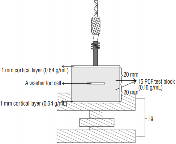
Fresh human cadaveric bone has been regarded as the most suitable medium for in vitro biomechanical bone screw fixation studies, but recent studies have shown that the density of cancellous bone can be highly variable1,4,5). In order to eliminate the variability in geometry and mechanical properties associated with cadaveric specimens, solid rigid polyurethane foam, which is comparable to human osteoporotic cancellous bone, can be used as an alternative test material for comparative evaluation of medical devices7,16,17,20). Therefore, in the present study, in order to have uniform samples for comparison, rigid polyurethane foam blocks (Sawbones, Vashon, WA, USA) were used as the test materials for the insertion of the screws. The foam had density of 15 per cubic foot (PCF) (0.16 g/mL), which has been shown to represent the material properties of osteopenic human cancellous bone4,24). Each 40├Ś40├Ś40 mm synthetic bone model was cut precisely into two 20 mm thick segments by cutting machine (JSG-520MB; JIN SAN T&G, Incheon, Korea), and a 1-mm layer of denser foam (0.64 g/mL) was laminated to the top and bottom surfaces to represent cortical bone. Fig. 2 shows the test setup. A custom testing jig was used to hold the blocks and prevent rotation during screw insertion. An ultra-thin load cell sensor was employed to minimize the displacement of the simulated fractures, and a 10-mm hole between 2 thin metal washers was placed at the center of the sensing head in order to accommodate the screw. A washer load cell (FlexiForce A201-100; Tekscan, Boston, MA, USA) was placed between the two foam block segments to measure the compression force and was then connected to a data acquisition system (MSO4034; Tektronix, Beaverton, OR, USA).
Each screw was prepared with two different lengths, 40-mm and 45-mm. We compared inter-fragmentary compression pressures between 40- and 45-mm long 4.5-mm Herbert screws (n=8 and n=9, respectively) and 40- and 45-mm long 4.0-mm cannulated lag screws (n=7 and n=10, respectively) after insertion into rigid polyurethane foam test blocks.
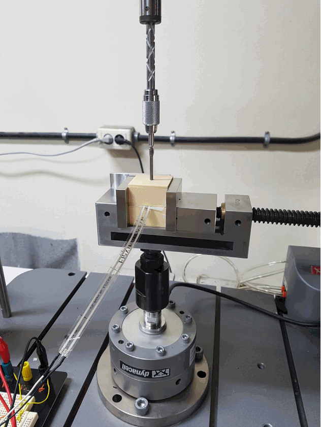
Because the total length of the foam block was 42 mm, we inserted the 40-mm screw within the cancellous foam block, but were able to penetrate the denser bottom cortical foam with the 45-mm screws. This allowed us to compare inter-fragmentary compression pressures as they are affected by penetration of the apical dens tip by the screws. All of the screws were placed according to the respective manufacturer guidelines and the specimen foam was drilled with drill bits specified by each screwŌĆÖs manufacturer. The screws were inserted at a controlled speed of 10 rpm until the threads were totally embedded in the foam. The test specimens were then mounted onto an universal testing machine (Instron3000; Instron, Norwood, MA, USA) (Fig. 3) and inter-fragmentary compression was produced by tightening the head of the screw to the surface of the first segment. Output from the washer load cells was measured during insertion of the screws, and the measurement continued after a steady state had been reached.
RESULTS
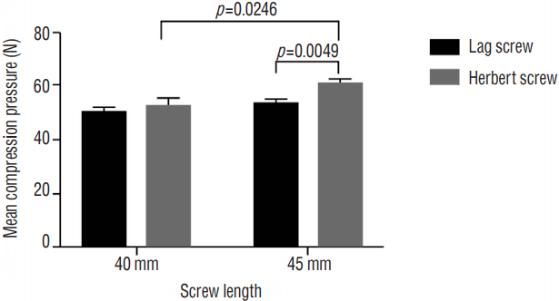
We compared the inter-fragmentary compression pressures between 4.5-mm diameter Herbert screws and 4.0-mm diameter cannulated lag screws with different lengths (40 mm and 45 mm). Mean compression pressure of the 40- and 45-mm cannulated lag screws were 50.48┬▒1.20 N and 53.88┬▒1.02 N, respectively, which did not reach statistical significance (p=0.0551). Mean peak compression pressure of the 40-mm long Herbert screw was 52.82┬▒2.17, which was not statistically significant compared with the 40-mm and 45-mm long cannulated lag screw (p=0.3679 and p=0.2473, respectively). However the 45-mm long Herbert screw had significantly greater mean compression pressure (60.68┬▒2.03 N) than both the 45-mm long cannulated lag screw and the 40-mm Herbert screw (p=0.0049 and p=0.0246, respectively) (Fig. 4).
DISCUSSION
The cannulated lag screws with a short thread are most frequently used for anterior odontoid fracture fixation to produce a lag effect across the fracture site2,6,9,13,20,21). Theoretically, after the cannulated lag screw has crossed the fracture line, the threads engage the fragment and the lag effect of the screw reduces the fracture. Further tightening of the lag screws pulls the odontoid in a caudal direction, compressing the fracture site and enhancing fracture stability, which results in improved healing of the fracture3,11,15,20,25).
A single piece non-cannulated headless compression Herbert screw with variable pitches of the proximal and distal threaded portions, was first designed by Herbert and Fisher18) in 1984, to provide internal compression and stability of scaphoid fracture while avoiding any protrusion of metal on the joint surface of the bone. And thereafter Whipple modified the Herbert screw by developing a cannulated version to allow for more accurate screw placement. Because of its security of fixation and lack of a protrusive head, the Herbert screw would appear to have significant advantages over standard implants for the fixation of small cancellous bone fragments. In 1994, Chang et al.8) first described the Herbert-Whipple screw fixation of type II odontoid fracture. They suggested that close reduction and compressive osteosynthesis by double-threaded compression screw is an optimal method of treatment of displaced type II odontoid fractures.
At our institute, we originally used the cannulated lag screw for anterior odontoid screw fixation, but recently, in accordance with the method introduced by Chang et al.8), we have adopted the 4.5-mm Herbert screw (Zimmer)10,11,19). In our previous study using the Herbert screw for anterior odontoid fixation, the risk of fusion failure was 21 times greater in patients with a fracture gap of more than 2 mm11). We believed that in order to facilitate maximal reduction of the fracture gap using the Herbert screw, it is essential for the screw to penetrate the apical dens tip, which has generally been the recommended surgical technique2,3). However, in fact, cortical purchase by the screw may be technically difficult and very stressful to the surgeon due to the risk of injury of adjacent basilar artery and pons3,25).
To date, there have been a number of many biomechanical studies that have evaluated different compression screws for bone fractures4,5,16,17,24). Burkhart et al.7) reported that there were no significant differences in the stability provided by a 3.0-mm headless compression screw and a 2.0-mm cortical screw in radial head fractures in fresh frozen human cadaveric bone specimen. A recent cadaveric scaphoid study found that the mean inter-fragmentary compression generated by an Acutrak 2 Standard screw was significantly greater than that of a Synthes headless compression screw16). However, there are few published biomechanical studies for odontoid screws. Magee et al.20) have reported a biomechanical comparison of a fully threaded, variable pitch screw and a partially threaded lag screw for internal fixation of type II dens fractures in human cadaveric specimens. They found that stiffness and load to failure were greater for the headless, fully threaded variable pitch screw compared with the partially threaded lag screw. However, they did not measure the inter-fragmentary compression pressure between the fracture fragments of the odontoid process.
Previous studies have shown that the density and elastic modulus of cancellous bone (e.g., scaphoid bone) are highly variable, and the mechanical properties of trabecular bone from multiple locations have tremendous variations, which can affect the maximum achievable compression force1,4,14). Rigid polyurethane foam is been regarded as an ideal material for in vitro evaluation of compression screws. The uniform and consistent properties of rigid polyurethane test blocks in comparison to animal and human bone has been well established. Foam densities of 10, 15, and 20 pounds PCF have been shown to have the material properties of osteoporotic, osteopenic, and normal bone, respectively4,17,24). Because type II odontoid fractures are the most common type of axis fracture in elderly patients, in the present study, we used 15 PCF foam (0.16 g/mL) in order to represent the material properties of osteopenic human cancellous bone.
In the present study for the 40 mm-long Herbert screw and the 40-mm long cannulated lag screw, there was no difference in inter-fragmentary compression pressures. But, in our comparison of 45-mm long screws, which were long enough to penetrate the dense cortical foam of the opposite test block, the 45-mm Herbert screw produced significantly higher inter-fragmentary compression pressure than the 40-mm Herbert screw. The 45-mm cannulated lag screws also provided greater inter-fragmentary compression pressure. Although this did not reach statistical significance (p=0.0551), we surmised that the number of test screws (n=7) was too small to achieve a statistical difference. These results support the recommendations of previous studies that, in order to facilitate maximal reduction of the fracture gap using lag screws, it is essential to penetrate the apical dens tip with the screw2,3,11).
When we compared the 45-mm Herbert screw and the 45-mm cannulated lag screw, the Herbert screw produced significantly larger peak compression pressures than the cannulated lag screws (p=0.0049). This result suggests that the specially designed Herbert screw may provide greater reduction of the fracture gap in the odontoid process than the cannulated lag screws when the screws penetrate the apical cortex of the dens. This finding further emphasizes the importance of screw-tip penetration of the apical dens tip during anterior odontoid screw fixation.
We have some limitations in this study. First, we did not check other biomechanical parameters, including bending strength or pullout strength. Inter-fragmentary compression pressure alone cannot guarantee the biomechanical superiority of the Herbert screw compared with the cannulated lag screw. Second, clinically, greater inter-fragmentary compression pressure may not mean greater reduction of the fracture gap in odontoid process fixation, because, in humans, the dens is attached by many strong ligaments including apical and alar ligaments12,26). Furthermore, because there are so many risk factors of fusion failure of odontoid process fixation2,3,11,15), we must consider other factors for higher bone fusion rates after odontoid screw fixation.
Third, in human, due to the heterogeneous fracture line and orientation of type II odontoid fracture, odontoid screw insertion generally have a little angled path against the fracture line during the operation. But, in this biomechanical study, we inserted the screws perpendicularly into the test blocks, which may not reflect the real situation of anterior odontoid screw fixation.
CONCLUSION
Our results showed that inter-fragmentary compression pressures of the Herbert screw were significantly increased when the screw tip penetrated the opposite dens cortical foam. This can support the generally recommended surgical technique that, in order to facilitate maximal reduction of the fracture gap using anterior odontoid screws, it is essential to penetrate the apical dens tip with the screw.


