INTRODUCTION
Retained foreign body granulomas in the intervertebral disc space due to penetrant traumas are very rare like other paraspinal ones2,3,5-7,10). To our knowledge, all of the reported retained foreign bodies in the intervertebral disc space were from missile injuries and there was not any report involving a retained foreign body granuloma in this region from wooden penetrant trauma2,3,5,6,10).
CASE REPORT
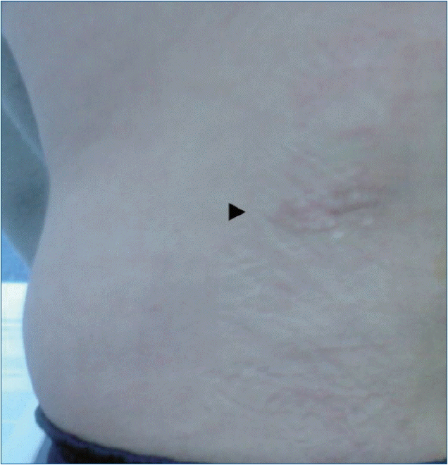
A 30-year-old man sought medical care with a history of low back pain (LBP) that radiated to lower extremities. He reported that he had fallen from a tree and a tree branch had been penetrated into him left lower back before 17 years. That tree branch had been taken out manually and he had recovered without any complaint. In the past two months, the patient has presented a LBP radiating to his lower limbs. Physical examination revealed a bad-healed scar at about level of the third lumbar vertebra on left paraspinal region (Fig. 1). He had pain on lower back with palpation. The characterization of the pain in his lower extremities was a dermatomal pattern. The straight-leg-raising sign was negative and there was not any neurologic deficit on lower limbs.
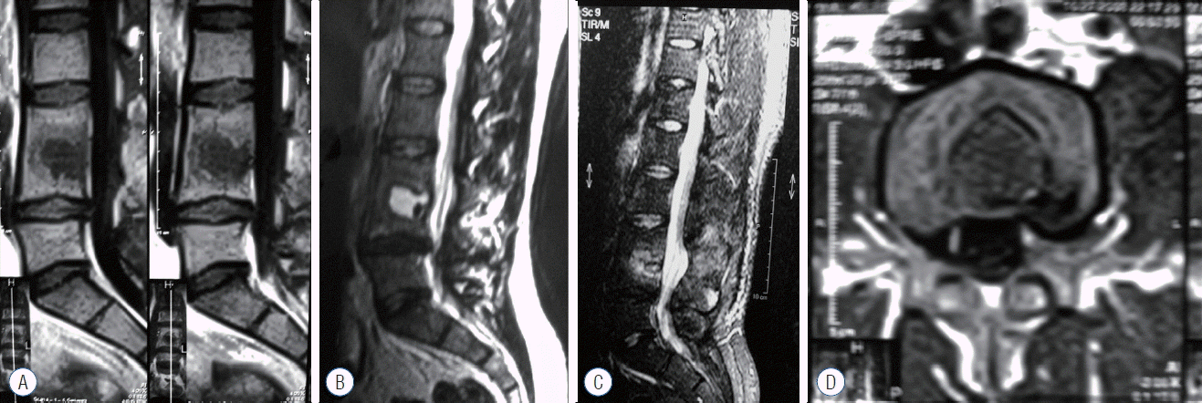
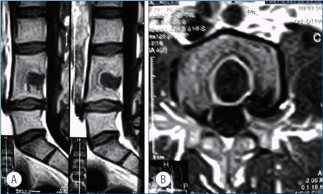
Computerized tomography (CT) demonstrated a hypodenselytic lesion in the L4 vertebral body surrounding a linear hyperdense structure (Fig. 2). Anterior part of the disc space between fourth and fifth lumbar vertebrae had disappeared with fusion of these bodies in magnetic resonance (MR) images. MR images also revealed a lesion that lodged in posterior part of this disc space and expanding into the corpuses of these vertebrae (especially into L4 vertebral corpus). This lesion was hypointense in T1- (Fig. 3A, D) and hyperintense in T2-weighted (Fig. 3B) and STIR sequences (Fig. 3C) images. It was seen a linear horizontal area of decreased signal intensity representing the intranuclear cleft in the center of posterior part of disc space between fourth and fifth lumbar vertebrae in sagittal MRI image of the STIR sequence. The peripheral areas of the lesion were enhanced after gadolinium injection (Fig. 4).
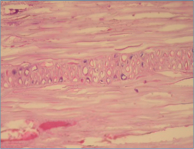
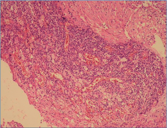
The patient underwent L4-5 hemilaminectomy via a midline skin incision. There was not any adhesion in the extradural space and around the dura. A firm mass was found lodged in posterior part of the intervertebral disc space between the fourth and fifth lumbar vertebrae. There was not any epidural expansion of the mass. Excision of the mass revealed a tough granulation tissue surrounding a tiny piece of wood in a little cavity filled with a clear yellowish fluid (Fig. 5). The mass was totally removed. There was a fusion between anterior parts of third and fourth lumbar vertebrae and a degeneration in the discal material in the rest of intervertebral disc space. Chronic granulation tissue and a wooden splinter were confirmed histologically (Fig. 6, 7). Postoperatively, the pain resolved gradually and the patient was discharged without any problem 10 days after operation. He had a total symptomatic cure after two months from excision of the foreign body.
DISCUSSION
Biological responses of tissues to foreign bodies are influenced by the location and the nature of them1-7,10). In a soft tissue, a wooden foreign body can cause either a mild inflammatory tissue reaction resulting in a localized granuloma or a severe reaction resulting in a significant infectious lesion with or without osseous reaction in adjacent bone1,4,6,8).
Paraspinal disorders like ones in the intervertebral disc space may cause important complications because they may affect vital structures in and near the spinal canal. In a similar way, the majority of reported cases with foreign bodies that embedded and retained near the spinal canal presented with delayed symptoms or findings resulting from compression of neural elements due to developed granuloma or abscess as well as in creased bony growths or migration of foreign body in the spinal canal2,3,5-7,10,11). In the literature, there are only four case reports presented with the retained foreign body granulomas in the intervertebral disc space from penetrant traumas2,5,6,8,10). All of them were from missile injuries mentioned previously. Two of these cases presented with cauda equina syndrome resulted from compression of neural structures due to granuloma and/or bony growths while two of them presented with symptoms and findings other than neurological deficits like our case2,5,6,8,10).
We thought that our patient’s complaints probably were due to expanding of granuloma into the corpora of adjacent vertebrae. His complaints totally disappeared after removing the granuloma that in a way supported our thought.
The detection of wooden foreign bodies with plain radiographies and CT has limited value because they are radiolucent. Therefore, ultrasound and MR imaging are recommended in the detection of these foreign bodies like of other nonmetallic ones9). But, the identification of wooden foreign bodies may also be difficult on MR imaging when they are small and on ultrasound when they are in the deeper paraspinal tissues. Thus, a wooden foreign body in paraspinal tissues like intervertebral disc space may be easily missed especially in acute cases. More over, it is very difficult to predict the natural history of a retained foreign body in the intervertebral disc space, because, a sufficient number of cases is unfortunately not exist in the literature.
CONCLUSION
If a retained wooden body in intervertebral disc space is missed and it is not removed, it may be potentially hazardous and result in significant morbidity because of the nature of wood and the paraspinal location of intervertebral disc space. Moreover, a missed foreign body in a paraspinal tissues may cause further damages for patients and malpractice claims for physicians. Therefore, physicians and radiologist need to keep their diagnostic suspicions height in terms of retained foreign bodies during examination or follow up a patient with paraspinal wooden penetrant traumas.