|
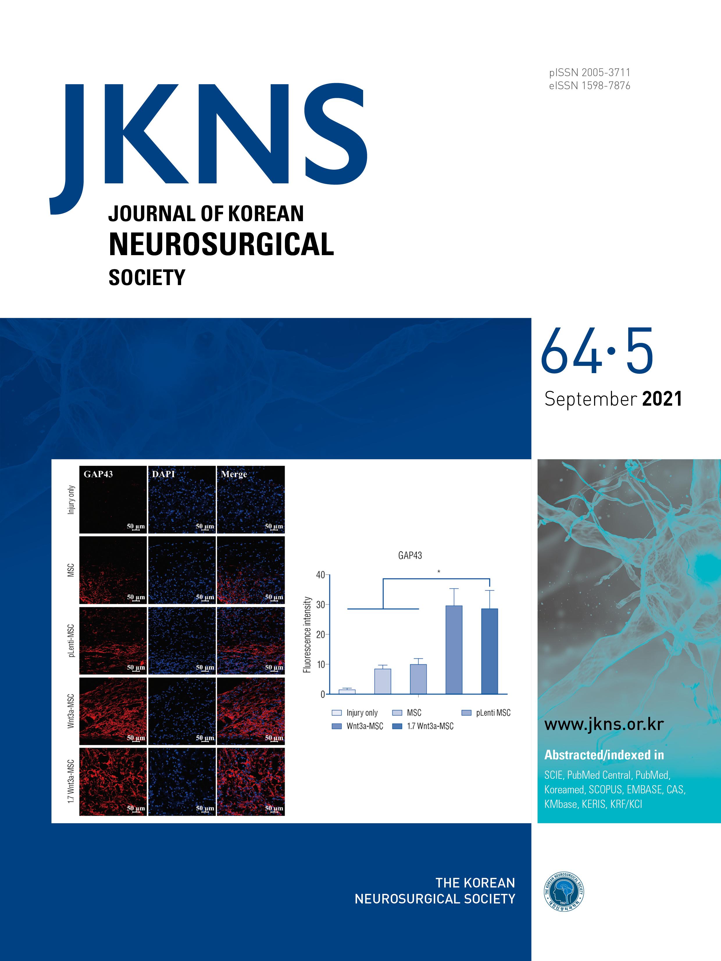
Immunofluorescent staining of axonal regeneration marker growth-associated protein 43 (GAP43). Confocal microscopic images revealed that anti-GAP43 antibody staining in the spinal cord was greater in the Wnt3a-MSC and 1.7 Wnt3a-MSC groups than in other groups. Tile scan image on left side, quantification of fluorescence intensity on right side.
|