|
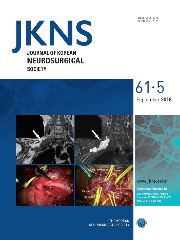
Brachial plexus magnetic resonance imaging revealed a well-defined contrast enhancing multilobulated large mass from C7 nerve root to middle trunk (white arrow). Coronal T1 weighted image shows the intermediate signal of brachial plexus tumor. Coronal T2 weighted short tau inversion recovery (STIR) image shows the high signal of brachial plexus tumor.
Intraoperative images of brachial plexus tumor removal shows that brachial plexus tumor is dissected. After the brachial plexus tumor removal, entire brachial plexus anatomy can be seen.
|