|
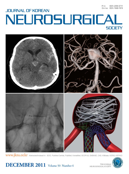
"Upper left : Initial brain computed tomography (CT) showing spontaneous
subarachnoid hemorrhage in basal and prepontine cistern. Upper right : 3D angiogram shows a broad neck basilar top aneurysm
involving both P1 segments and basilar artery. Lower left : Unsubstraction
image showing 4.5 mm×28 mm Enterprise stent placed in the distal basilar artery and proximal left PCA and 4.5 mm×20 mm Neuroform stent is deployed into aneurysm sac. Lower right : 3D computer illustration graph shows a Enterprise stent placed in the distal basilar artery and proximal left PCA and Neuroform stent is located into aneurysm neck."
|