|
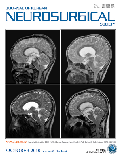
Midsagittal brain MR images showing the cervicomedullary junction and high cervical spine obtained in a 15-year-old girl presenting with neck pain. Note the nine-year gradual progression of cerebellar tonsillar herniation from 7 mm (upper left) to 24 mm (lower right), the descent of the obex from 2 mm above the foramen magnum (upper left) to 8 mm below (lower right) and the thinning of the medulla oblongata from 8 mm (upper left) to 6 mm (lower right). Upper left : Initial sagittal T2-weighted MR image. Upper right : 2 years. Lower left : 7 years. Lower right : 9 years after the initial presentation.
|