INTRODUCTION
Cranial irradiation has been widely used as a therapeutic method treating a wide variety of lesions, particularly neoplasms. Though radiation therapy of central nervous system is usually well-tolerated, it does occasionally cause clinically significant long-term toxicity such as late delayed radiation necrosis and irradiation-related arteriopathy with stroke14). Among other complications induction of neoplasm is a rare but well-documented serious sequela of therapeutic irradiation. Radiosurgery is not free from this problem although there are only a few reported cases14). We report a case of glioblastoma which was developed after gamma knife radiosurgery (GKS) for meningioma.
CASE REPORT
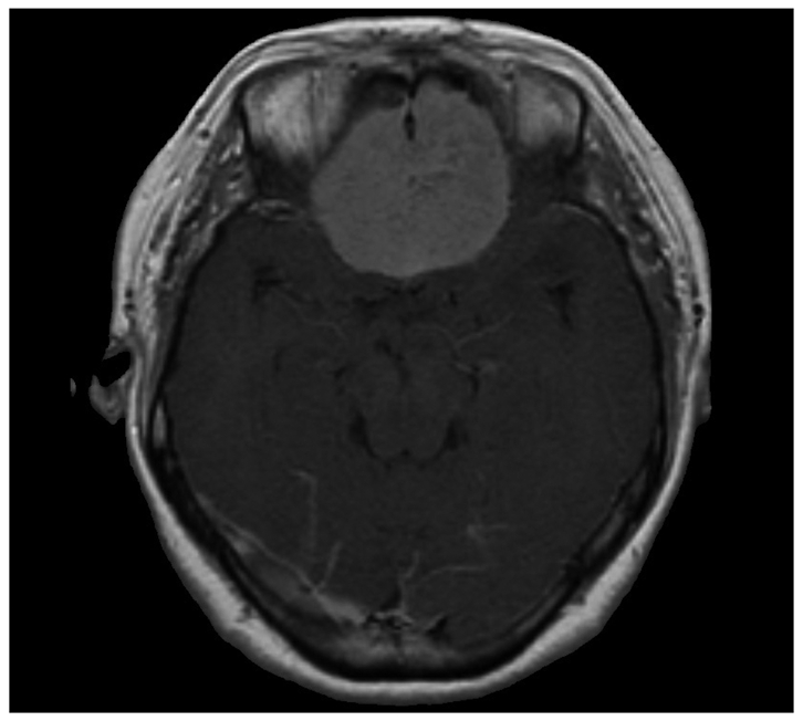
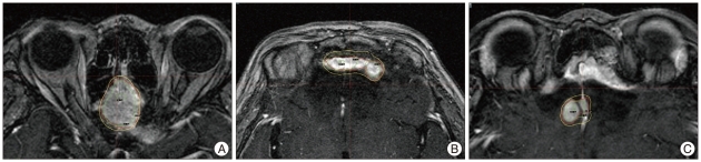
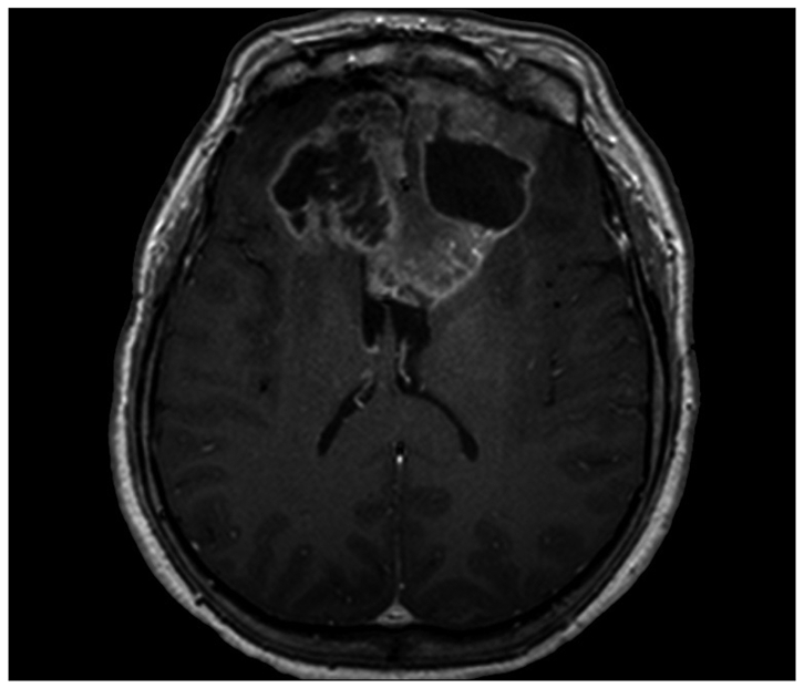
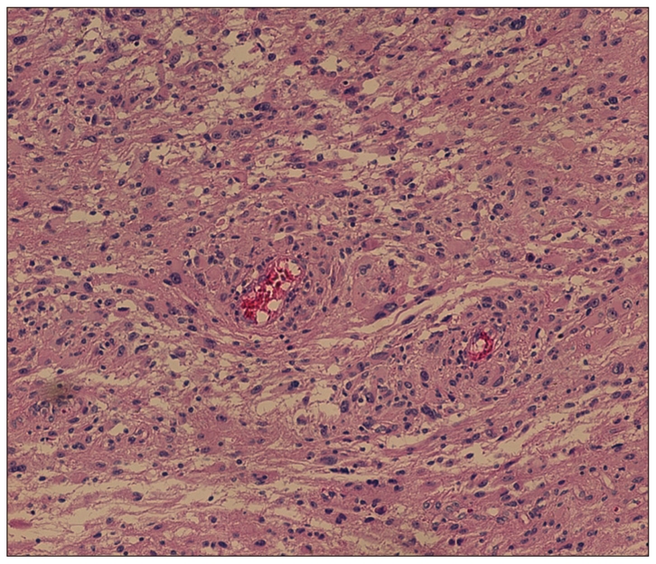
A 47-year-old woman presented with headache, anosmia, and visual dimness. Large extra-axial mass originating from olfactory groove and both frontal base was found on MRI (Fig. 1). She underwent gross total tumor removal, and the pathological diagnosis of meningotheliomatous meningioma was confirmed. Approximately 1 year after surgery, the follow-up MRI scan revealed tumor recurrence. She underwent GKS with 16 Gy of marginal dose at 50% isodose line (Fig. 2A). Three years after initial radiosurgery, the subsequent MRI scan revealed tumor progression at a location different from the previous recurrence. She underwent radiosurgery with 13 Gy of marginal dose and she was subjected to third GKS with 15 Gy of marginal dose on local recurrent lesion after 2 years from second GKS (Fig. 2B, C). At 58 months from the initial GKS, which is also 72 months from the first operation the patient experienced severe headache with vomiting and a huge new lesion was found in the area of the previous radiosurgery (Fig. 3). She underwent the second craniotomy and tumor removal. On this occasion, the pathological finding was consistent with glioblastoma (Fig. 4). She was treated with concomitant chemo-radiotherapy (total dose of irradiation 6000 cGy over 30 fractions and oral temozolomide 75 mg/m2/day during the radiotherapy period) followed by adjuvant chemotherapy with oral temozolomide. First cycle was consisted of 150 mg/m2/day temozolomide for 5 days, and subsequent 5 cycles were maintained with 200 mg/m2/day for 5 days with 4 weeks interval between each cycle. The tumor progression was significant at 11 months from the diagnosis of glioblastoma with neurological deterioration. Ventriculoperitoneal shunt was done for progressive hydrocephalus. Currently, she is alive at the time of this writing (15 months after diagnosis of glioblastoma) with only supportive management.
DISCUSSION
There is now increasing concern regarding the potential for radiation-induced neoplasm, because of the intensive use of radiotherapy under a wide range of doses and treatment conditions resulting in a longer survival of brain tumor patients2,14,24). Despite the reported documents about the safety of radiosurgery, additional reports of glioma induction after radiosurgery may temper its use in the treatment of benign lesions, such as meningiomas, particularly in younger patients10,12). The following criteria for radiation-induced neoplasms have been previously described7,11) : 1) a second tumor occurs within the field of irradiation used to treat the primary disease; 2) the tumor is not present prior to irradiation, and there has been a reasonable interval between radiotherapy and the detection of the second tumor (usually several years); 3) a histological difference exists between the primary and subsequent tumor; and 4) no known genetic or predisposing conditions to secondary malignancy are observed. In other series, the criterion of latent period in radiation induced tumor was set to 5 years5,6,13). However, the "5 year" period is not accepted as an absolute standard due to insufficient biologic evidence. Therefore, the present case could be considered as radiation induced tumor. In this way, the present case fits the all criteria and deserves to be diagnosed as radiation-induced malignancy.
The mechanism of radiation-induced neoplasms is multifaceted and not well understood. However, radiation is now well recognized to be a trigger, not only a treatment, for cancer. When the cells are irradiated, the probability of malignancy increases with dose, most likely with no threshold. That is, no dose is too small to be effective. This view is based on experimental data showing that even a single photon can cause a base change leading to mutations that eventually can cause malignancy. In contrast, the severity of malignancy is not dose related. That is, a cancer induced by a small dose of radiation is not less harmful than a cancer induced by a large dose. Furthermore, the absolute value of radiation dose in stereotactic radiosurgery is low. But, in the concept of biological equivalent dose, it is not a low-dose treatment, thus it could cause radiation induced tumor. The lower incidence of radiation induced tumor in radiosurgery, could be explained by relatively smaller volume of normal tissue exposed to radiation compared to conventional radiation. Brada et al.4) reported the risk of second brain tumor formation in 334 patients treated for pituitary tumors with surgery and fractionated small-field irradiation (median dose, 45 Gy). With a cumulative follow-up period of 3760 years, five patients developed second tumors (two astrocytomas, two meningiomas, and one meningeal sarcoma). The latency period is similar to what has been seen with fractionated therapy in the several years period after radiosurgery, and malignant tumors occur earlier than their benign tumor counterparts. The cumulative risk of developing second brain tumors was 1.3% at 10 years and 1.9% at 20 years. The relative risk of a second brain tumor was 9.38 compared with the normal population4).
Concerning the risk of developing a secondary neoplasm, several factors such as patient age, tissue vulnerability, radiation type and dose, underlying disease, and additional chemotherapy may all play an important role2,4,17,24,25). The relative risk of a secondary tumor following cranial irradiation in children has been estimated to be from 2.6 to 38.8 compared with the risk in the standard population8,15,22) while in the adult population it was estimated to be from 9.38 to 164,24). In some cases, younger patients have been reported more vulnerable for second tumor development. Sadetzki et al.16) described 253 patients who developed meningiomas after radiation for tinea capitis. The mean time from exposure to meningioma diagnosis was 36 years (range, 12-49 year). The authors found a higher incidence of multiple lesions, a younger age at diagnosis, and a higher percentage of calvarial lesions than in control patients who developed meningiomas without previous exposure to ionizing radiation16).
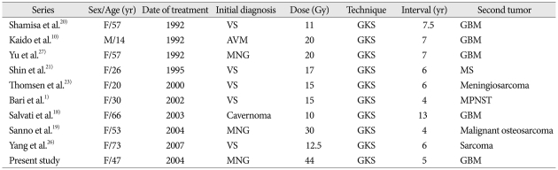
The toxicity of therapeutic doses of radiation is primarily acute. However, the carcinogenic effects are not restricted to those tissues manifesting clinical toxicity. In fact, the opposite may be more accurate, because high dose of radiation may sterilize the carcinogenic potential of a tissue by killing cells. Only in those cells that are not killed deoxyribonucleic acid repair errors may lead to transforming mutations. The efficiency of tumor induction varies inversely with repair capacity, which in turn depends on the integrity of cell cycle check-points3,9). The general form of the dose-response curve for radiation associated second tumors is not clear, but several experiments on small animals suggest that the incidence increases with dose up to a maximum usually occurring between 3 and 10 Gy, followed by a subsequent monotonic decrease3). There are also studies reporting that the highest incidence of radiation associated second tumors occurs at field peripheries, where the dose is less than at the field center9). Most of the reported cases of radiation-associated tumor were related to conventional radiotherapy. However, malignant tumors after GKS of benign tumors have rarely been discussed. There were small number of malignant neoplasms after treatment of vestibular schwannoma, meningioma or arteriorvenous malformation (Table 1). Considering that carcinogenetic potential is not necessarily dose-dependent and sublethal damage and repair errors are required for development of secondary tumors, single session radiosurgery may be relatively safer than fractionated radiotherapy with repeated chance of sublethal damage. However, it seems that radiation-induced malignancy can occur within very low-dose peripheral regions as well as the full-dose regions. Because the number of the patients who underwent radiosurgery and long-term follow up is much smaller than that treated with fractionated radiotherapy, relative safety of radiosurgery needs to be proven by further accumulation of clinical data. We draw a conclusion that the number of GKS performed, is not important. As cumulative level of radiation increases by the repeated GKS, so does the oncogenic opportunity. In the present study, the cumulative radiation dose of three repeated GKS is higher than that of conventional radiotherapy, leading to oncogenic change. In our series, clinical outcome was not different and as poor as primary glioblastoma treated with standard treatment of surgery, radiotherapy and chemotherapy. Although malignancy after radiosurgery is very rare, its impact is not acceptable considering the benign nature of the primary disease. No one knows for sure that how much dose of the radiation causes oncogenic transformation. Therefore, the possibility of this fatal complication should be balanced always in case of radiosurgery.