| Home | E-Submission | Sitemap | Editorial Office |  
top_img
Journal of Korean Neurosurgical Society > Volume 65(3); 2022 > Article
Park: Complex Pathophysiology of Abusive Head Trauma with Poor Neurological Outcome in Infants

Abstract

Abusive head trauma (AHT) in infants, especially acute subdural hematoma, has an extremely poor outcome. The most decisive and important finding is the appearance of a widespread low-density area on head computed tomography. This phenomenon was traditionally thought to be caused by cerebral ischemia. However, many other pathophysiological abnormalities have been found to be intricately involved. Recent studies have found that status epilepticus and hyperperfusion injures are the major causes. Another serious problem associated with AHT is cardiopulmonary arrest (CPA). Many infants are reported to visit to the hospital with CPA, and its pathophysiology has not been fully elucidated. This paper examines the background of these pathological conditions and associated factors and elucidate the pathophysiological mechanisms resulting in poor outcomes in AHT. In addition to the intensity of assault on the head, the peculiar pathophysiological characteristics in infants, as well as the social background specific to child abuse, are found to be associated with poor outcome.

INTRODUCTION

Abusive head trauma (AHT) in infants, especially acute subdural hematoma, has an extremely poor outcome. Deaths are common in such cases, and even if the patient survives, severe neurological sequelae, such as delayed mental development, are expected [3,15,16,23,24,28,31,36]. The pathophysiological mechanisms resulting in more severe outcomes have not been fully elucidated. However, an extensive low-density area (eLDA) on head computed tomography (CT) is considered to be a crucial factor in exacerbating outcomes [14,17,18,42].
Traditionally, such mechanisms were believed to be ischemic changes, but it has now become clear that various pathological conditions are involved in a complex manner resulting in poor neurological outcomes [11,21,29].
In this paper, we discuss the pathophysiology of eLDA in AHT and the mechanism of cardiopulmonary arrest (CPA) that occurs after head trauma. We also discuss the decisions regarding AHT, that have recently caused major social turmoil [7,35,36].

WHAT IS THE BIG BLACK BRAIN (BBB)?

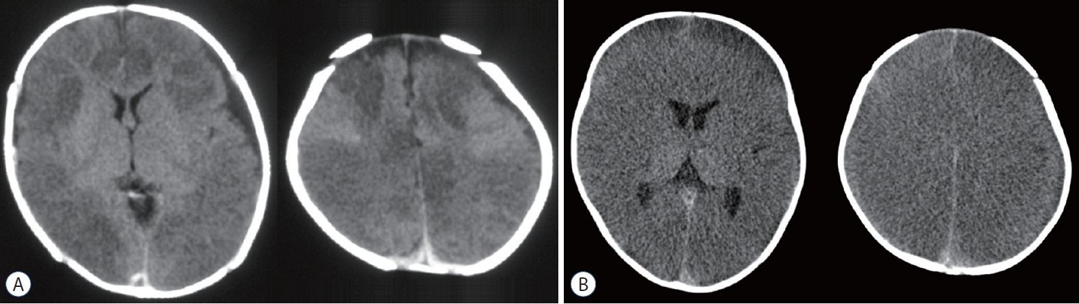
The BBB is a concept advocated by Duhaime et al. [14] and Duhaime and Durham [18]. They described the BBB as hemispheric hypodensity in head CT associated with acute subdural hematoma. This characteristic phenomenon is specific to head trauma in infants and does not occur in adults. It occurs in several AHT cases but can also occur in accident cases. Various physiological stresses, such as hypoxia, hypotension, hypercarbia, and seizures are involved as the onset factors, along with hypermetabolism and hyperpefusion. Recent animal studies have demonstrated that among the above mentioned factors, seizure duration is the most important [11].
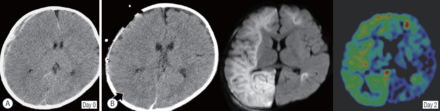
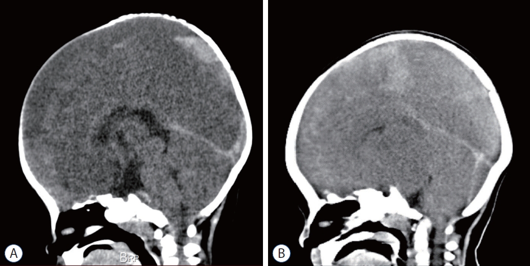
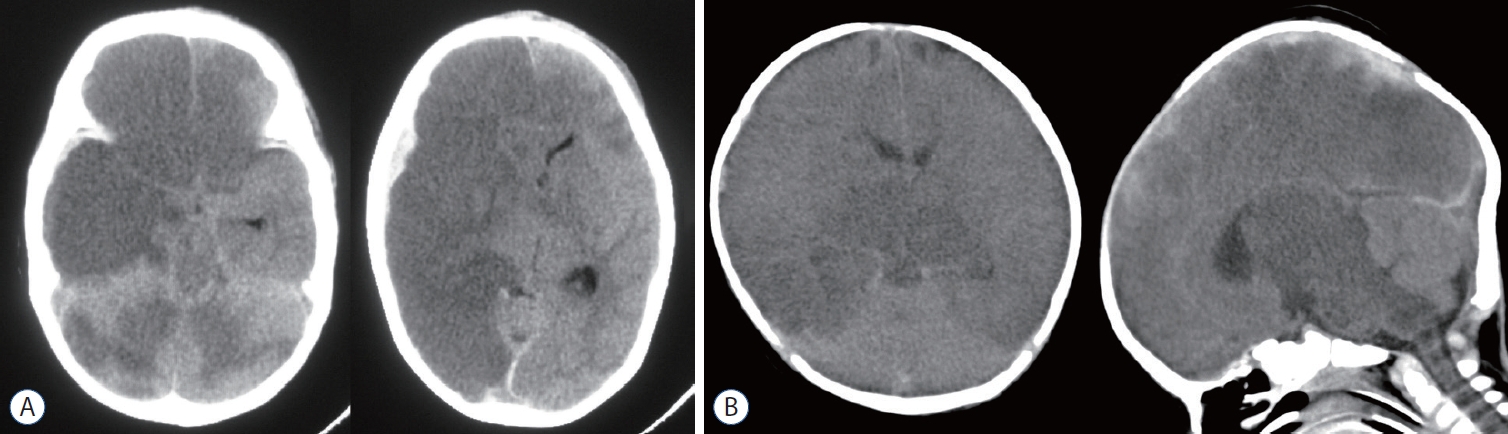
In the original report of the BBB, CT findings were typically characterized by lack of damage to the basal ganglia and thalamus (Fig. 1A) [14,17,18,21]. However, in actual clinical practice, there are many cases where the eLDA extends to the basal ganglia and thalamus (Fig. 1B). Another important fact is the time at which this finding is observed. The BBBs are commonly believed to occur hours to days after head injuries but is often observed on the first CT in some case.
While deepening our understanding of the concept of the BBB, we examined the eLDA pathology, which has a different background from that of the original BBB, and elucidated the cause of aggravation of AHT.

THREE PATTERNS OF extensive low-density area AND THEIR PATHOPHYSIOLOGY

Based on the findings on head CT, eLDA patterns are classified into the following three types according to three factors : when it occurs, where it occurs, and the presence of midline shift (MS).
Pattern A shows that eLDA has already been observed on the initial head CT, is widespread in bilateral cerebral hemispheres, and is not accompanied by MS. Pattern B implies that eLDA has already been observed on the initial head CT, is widely distributed in the unilateral cerebral hemisphere, and is accompanied by severe MS. Pattern C indicates that eLDA appears several days after the injury, may be local or extensive, and is often not accompanied by MS.
It is inappropriate to simply consider the condition that causes eLDA as a cerebral infarction. Other possible pathological conditions include respiratory disorders, brain swelling, status epilepticus (including non-convulsive) [5,26,27,37], hyperperfusion injury [35,38,45,46], arterial compression derived from cerebral herniation, and diffuse brain injury. In addition, there might be an unexplained pathophysiology in infants.
Here, we investigated the possible pathophysiological abnormalities in each pattern. The main pathological condition that results in pattern A is damage caused by respiratory distress, that is, hypoxic ischemic encephalopathy (HIE). Furthermore, in most severe cases, this condition is a typical finding in CPA. The main causes are direct respiratory center damage and traumatic shock associated with head injury, but choking due to vomiting is another cause to be noted. CPA caused by head trauma will be discussed in detail in the next chapter.
Pathological conditions associated with pattern B include status epilepticus and brain swelling immediately after the injury. Infants are more likely to have brain swelling than adults after head injuries. This is related to the effect of water content in the brain and response to hematomas. Finally, the pathology that results in pattern C includes status epilepticus as well as hyper(re)perfusion encephalopathy and mechanical compression of major arteries caused by cerebral herniation.
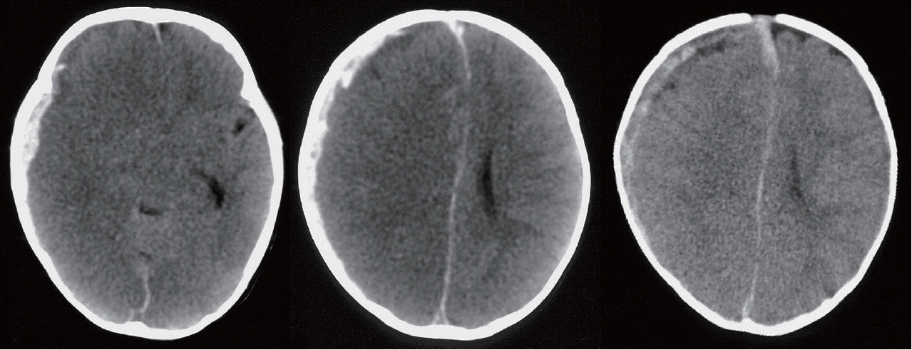
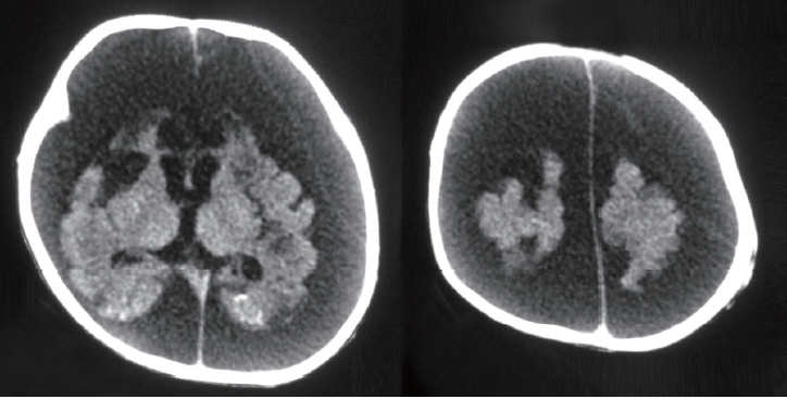
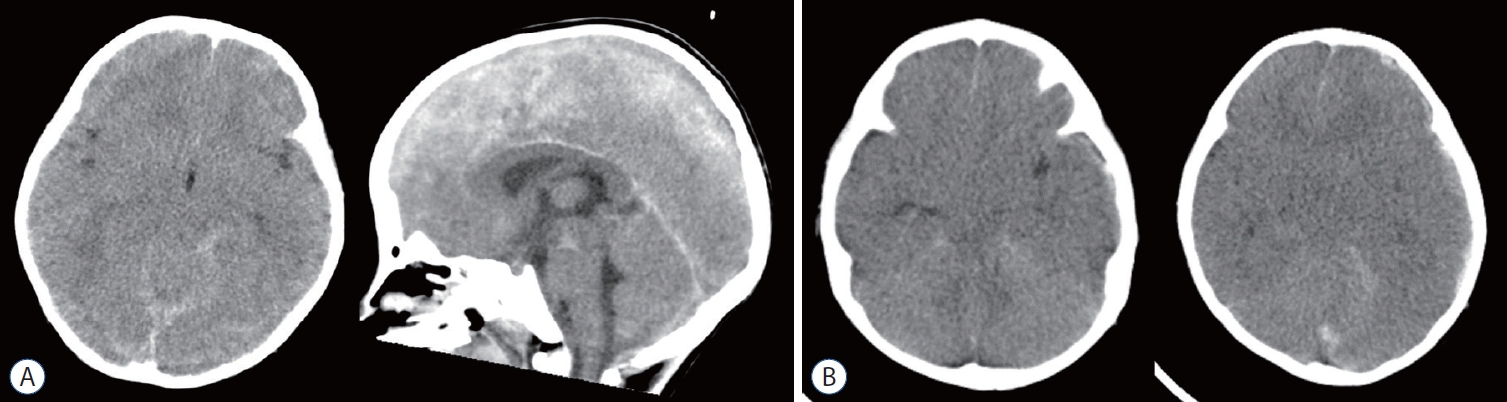
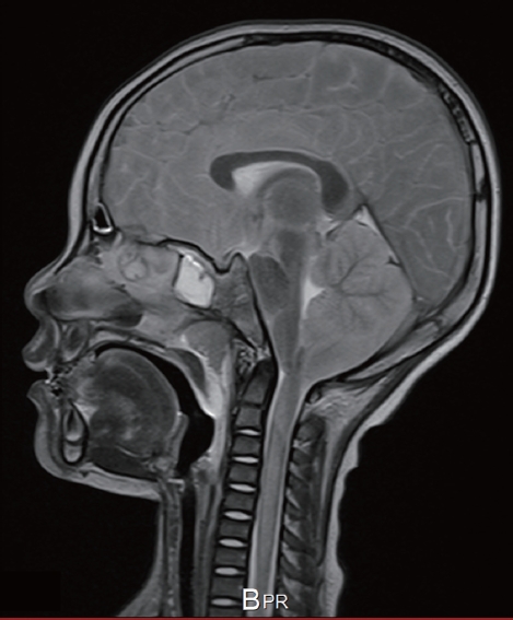
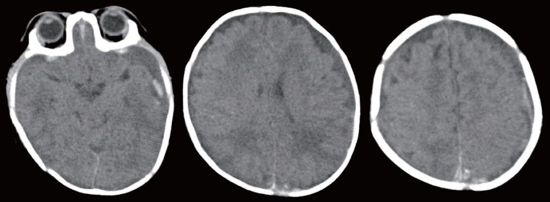
A typical case of each pattern is presented. A 5-month-old girl presented with CPA. Initial CT showed extensive hypodensity changes in the bilateral cerebral hemispheres. LDA extended to the basal ganglia and thalamus. Acute subdural hematoma was also observed in the left cerebral hemisphere, but this might not be the direct cause of CPA (pattern A) (Fig. 2). A 6-month-old girl was thrown on the floor by her father. She was admitted to the hospital because of impaired consciousness and status epilepticus. Head CT showed a right acute subdural hematoma with severe MS and widespread hypodensity in the right cerebral hemisphere (pattern B) (Fig. 3).
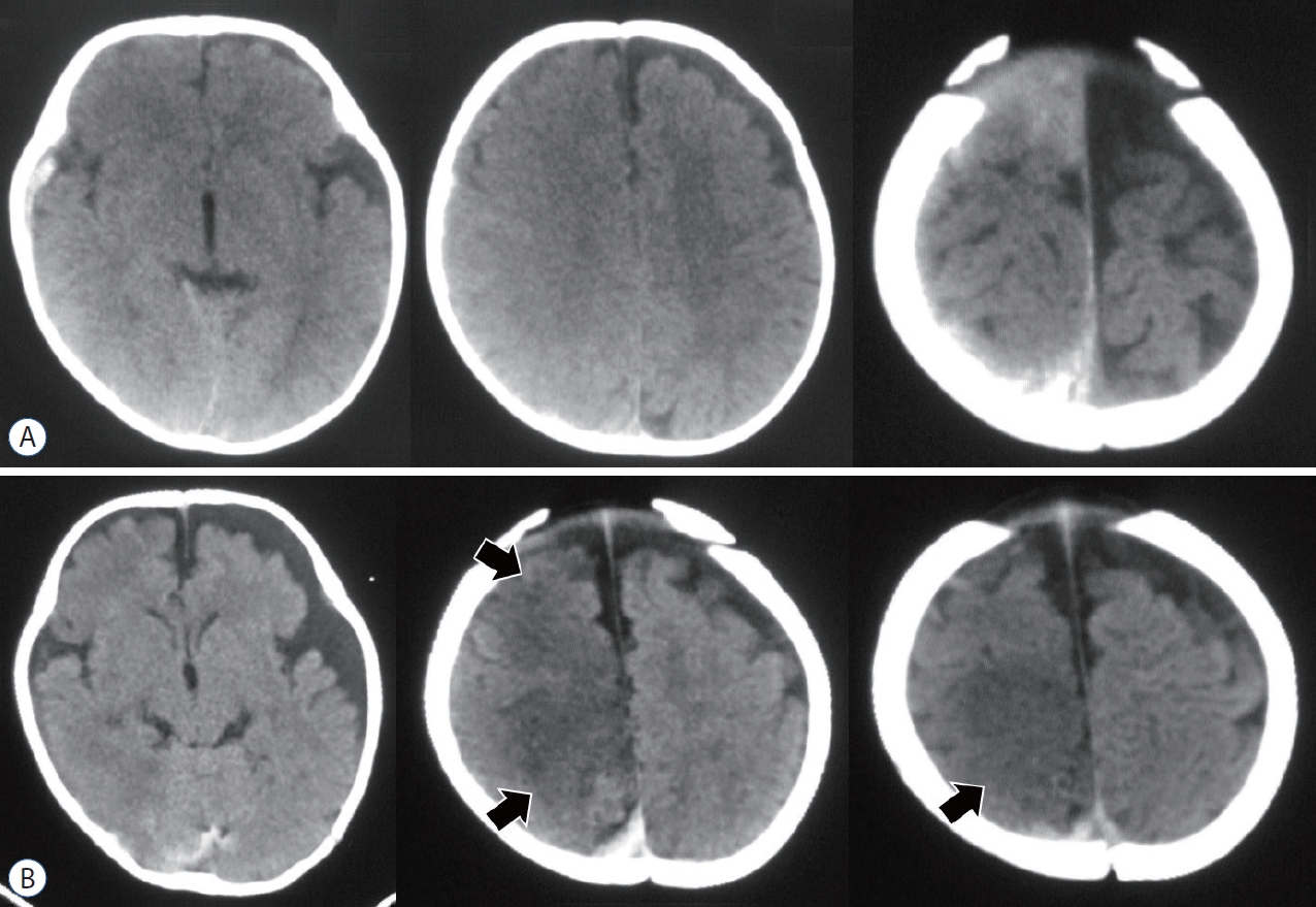
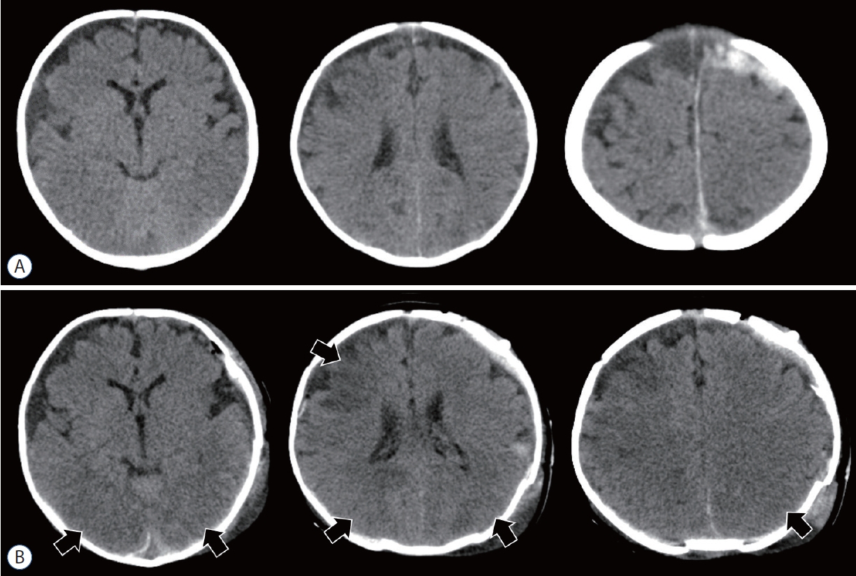
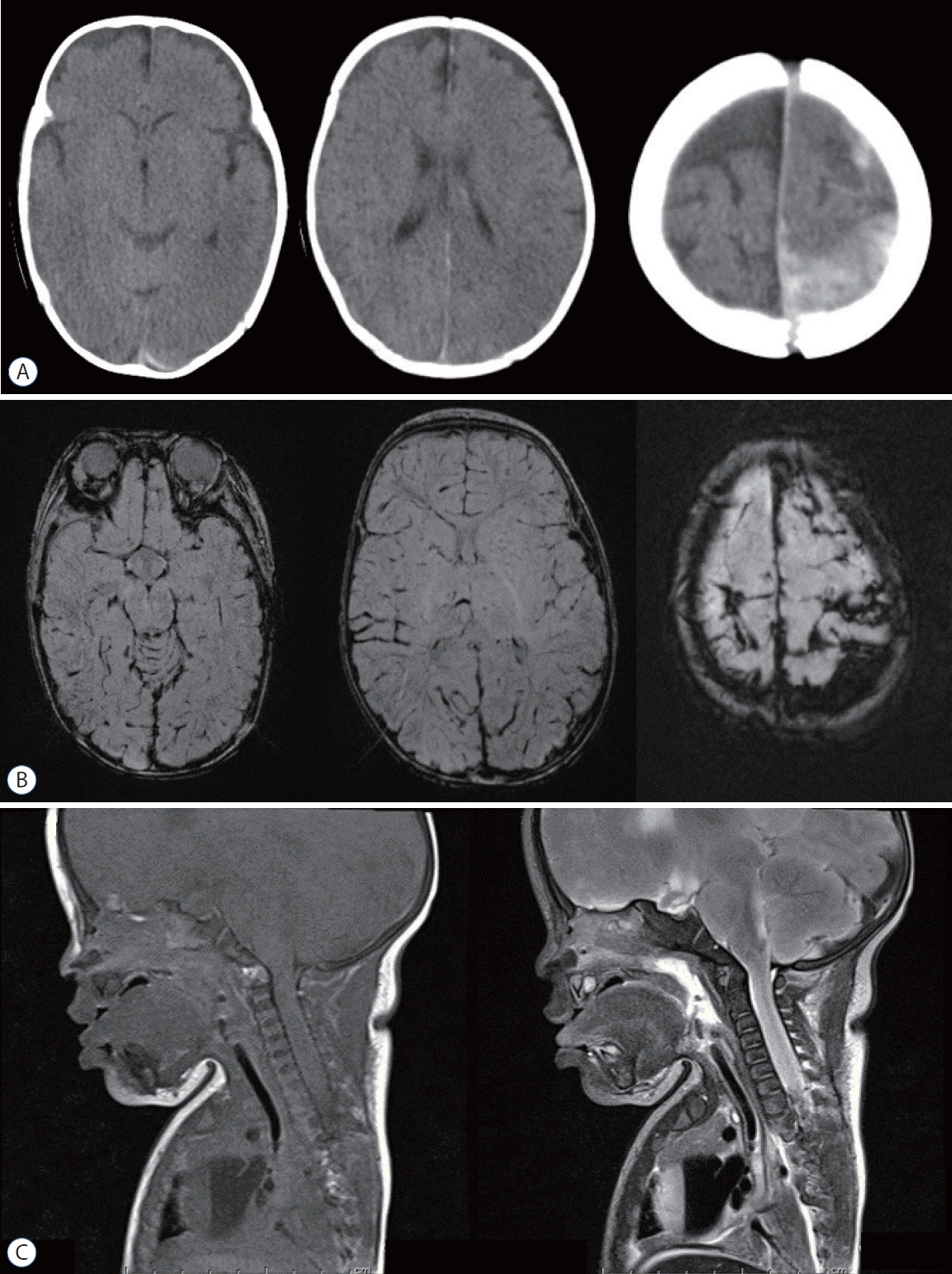
A 4-month-old boy was considered to have shaken baby syndrome (SBS) as the mechanism of injury. The infant was transported to another hospital with status epilepticus. eLDA in the right frontal parietal lobe, which was not visible on the initial CT, was revealed 3 days later (pattern C) (Fig. 4). A 3-month-old girl was also diagnosed with SBS. A craniotomy was performed for the acute subdural hematoma on the left side. Head CT performed 2 days after surgery revealed eLDA in the right frontal lobe and bilateral parietal lobes (pattern C) (Fig. 5).
As mentioned above, eLDA cannot be explained in thorough uniform pathophysiological abnormalities. In the case of AHT, HIE is considered to be an important pathophysiological abnormality, but status epilepticus (Fig. 4) and hyperperfusion encephalopathy after decompressive craniotomy (Figs. 5 and 6) should also be considered as major etiologies of eLDA. In other words, cerebral ischemia alone is not sufficient for explanation. However, once such eLDA is observed, rapid cerebral atrophy associated with cortical lamina necrosis occurs, and the prognosis of neurodevelopmental function is extremely poor (Fig. 7).

WHY DOES AHT CAUSE CPA?

Based on the results of clinical studies reported so far, there are four pathological conditions in which an infant's head injury can be exacerbated by CPA [39]. 1) Cerebral herniation (uncal herniation) : intracranial hematoma and brain swelling cause uncal herniation, and subsequent compression of the brain stem results in impaired consciousness and respiratory arrest. This pathophysiology is a common process from severe head injury to death and is not limited to AHT. 2) Diffuse brain injury : when a strong shearing force is applied to the head, it causes diffuse damage to the brain, including the brain stem, leading to severe consciousness disturbance immediately after the injury and respiratory arrest in the most severe cases. This involves the pathology of clinical diffuse axonal injury (DAI). 3) Injury to the respiratory center : when a direct external force is applied to the medulla oblongata or upper cervical cord, respiratory disorders can occur immediately afterwards. And 4) choking due to vomiting : infants are prone to vomiting after head trauma. The airways are blocked by vomitus, leading to respiratory arrest.
In clinical practice, owing to the presence of anterior fontanelle and unfused cranial sutures, head injuries in infants rarely exacerbate from cerebral herniation to CPA in a short period of time. Without careful observation and proper care within hours to days after injury, intracranial hematomas and cerebral swelling can be aggravated to the worst of CPA. Such cases are judged to be AHT, where the neglect factor is deeply involved.
Diffuse brain injury (clinical DAI) is difficult to diagnose using neuroimaging. Abuse experts have argued that there is extensive primary brain parenchymal injury, only in the presence of subdural hematomas and the presence of widespread LDA in the cerebral hemisphere [7,35,36]; however, this view might be inaccurate. The possibility of HIE due to asphyxia cannot be ruled out.
Therefore, what are the diagnostic imaging findings of primary diffuse brain injury? We must refer to the findings of DAI in diagnostic imaging that have been reported to date. The diagnostic imaging of DAI that occurs in adult cases should also be applied to infant cases. In most DAI cases, micro bleeding occurs in the deep cerebral white matter, corpus callosum, basal ganglia, brain stem, and lateral ventricle, and traumatic subarachnoid hemorrhage (T-SAH) in the basal cistern and peri mesencephalic cistern [1,20,32,33]. In some cases, acute diffuse brain swelling occurs, resulting in narrowing of the ventricular and cistern systems [10,44]. Head trauma experts believe that DAI occurs when these decisive findings are observed on the first head CT scan and causes a serious impairment of consciousness (Fig. 8).
In few cases, such findings cannot be observed. Thus, other etiologies should be considered. It can be said that this is the limitation of AHT diagnosis by neuroimaging (Fig. 9).
When the brain stem, especially the medulla oblongata, is directly damaged by head injury, not only consciousness disorder but also respiratory disorders occur immediately after the traumatic event, resulting in CPA in the worst-case scenario [19,30,34]. Traumatic primary brainstem injury should be evaluated using magnetic resonance imaging (MRI) findings. However, MRI examination is often dangerous in situations of unstable circular breathing. In rare cases, primary brainstem injury can be detected by CT, but in most cases, the artifacts associated with thick bony structures of the posterior fossa interfere with the diagnostic imaging of brainstem injury. Therefore, how should we diagnose primary injury of the medulla oblongata? The presence of hematoma on the posterior surface of the clivus can be an indirect finding of medulla oblongata injury (Fig. 10A). The other is the finding of an early edema with brainstem swelling (Fig. 10B). Brainstem damage derived from hypoxia or hypoperfusion does not cause swelling at an early stage.
Of course, it is the most useful rationale if an autopsy can prove traumatic primary damage to the medulla oblongata. This might include pathohistological diagnostic findings, such as DAI [2,22,43,47].
Upper cervical spinal cord injury is another important non-negligible factor that increases the severity of AHT [8,9,25]. To elucidate why CPA occurs, it is necessary to consider the possibility of upper spinal cord injury. In the case of a traffic accident or a fall accident from a high place, the golden rule of the initial treatment of trauma is to treat spinal cord injury regardless of age. In infants and children, a peculiar pathological condition may occur called spinal cord injury without evidence of radiologic abnormality (SCIWORA), which is a spinal cord injury without bone injury [6,40,41]. It might be easy to imagine the mechanism by which violent shaking causes severe SCIWORA and, in the worst-case scenario, respiratory arrest. However, one major blind spot is that only the findings of acute subdural hematoma and HIE in head CT are focused on and establishing the diagnosis of AHT is discussed, but the evaluation of upper cervical spinal cord injury is unfocused. It may be unavoidable that spinal cord MRI cannot be evaluated in CPA cases, but it is extremely problematic that spinal cord evaluation is often missed by close examination in autopsy. Retinal hemorrhage, multiple rib fractures, and long bone fractures are recognized as important extracranial injuries in AHT diagnosis, but ligamentous neck and/or spinal cord injury is also extremely crucial, especially in CPA cases (Fig. 11).
Additionally, the pathophysiology that must be kept in mind regarding the mechanism of CPA in infant head trauma is asphyxia due to vomiting. Vomiting occurs frequently in children with head injuries, especially in infants. Furthermore, infants have anatomical feature of a large tongue and a small trachea, and physiological feature of having more secretions in their laryngopharynx with a weak cough reflex. Regardless of the severity of the head injury, vomiting causes choking, and accidental injuries result in CPA in the worst-case scenario (Fig. 12). It is inaccurate to make an instant diagnosis of violent trauma solely based on a CPA case. Asphyxia due to vomiting can occur in either AHT or accident. Thus, additional evidence is required to judge AHT.
Of these four etiologies, diffuse brain injury and injury to the respiratory center can almost certainly be caused by AHT. Unless there are obvious assault assumed findings, such as multiple skull fractures or other physical injuries, it is important to note that the pathophysiology that resulted in CPA due to cerebral herniation or choking cannot be judged as definite AHT.
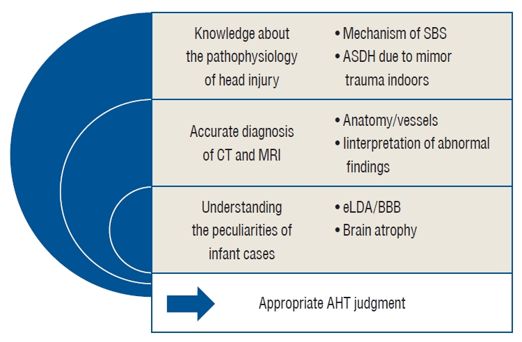
As mentioned above, it is not easy to determine whether a case of infantile acute subdural hematoma with CPA might be AHT. Appropriate and accurate diagnosis requires familiarity with head trauma and infant-specific anatomical and physiological characteristics (Fig. 13).

FACTORS THAT CAUSE AHT TO BECOME SEVERE AND RESULT IN POOR OUTCOME

Most AHT cases become severe and have poor outcomes, and it is necessary to consider various pathological conditions and backgrounds as etiological factors.
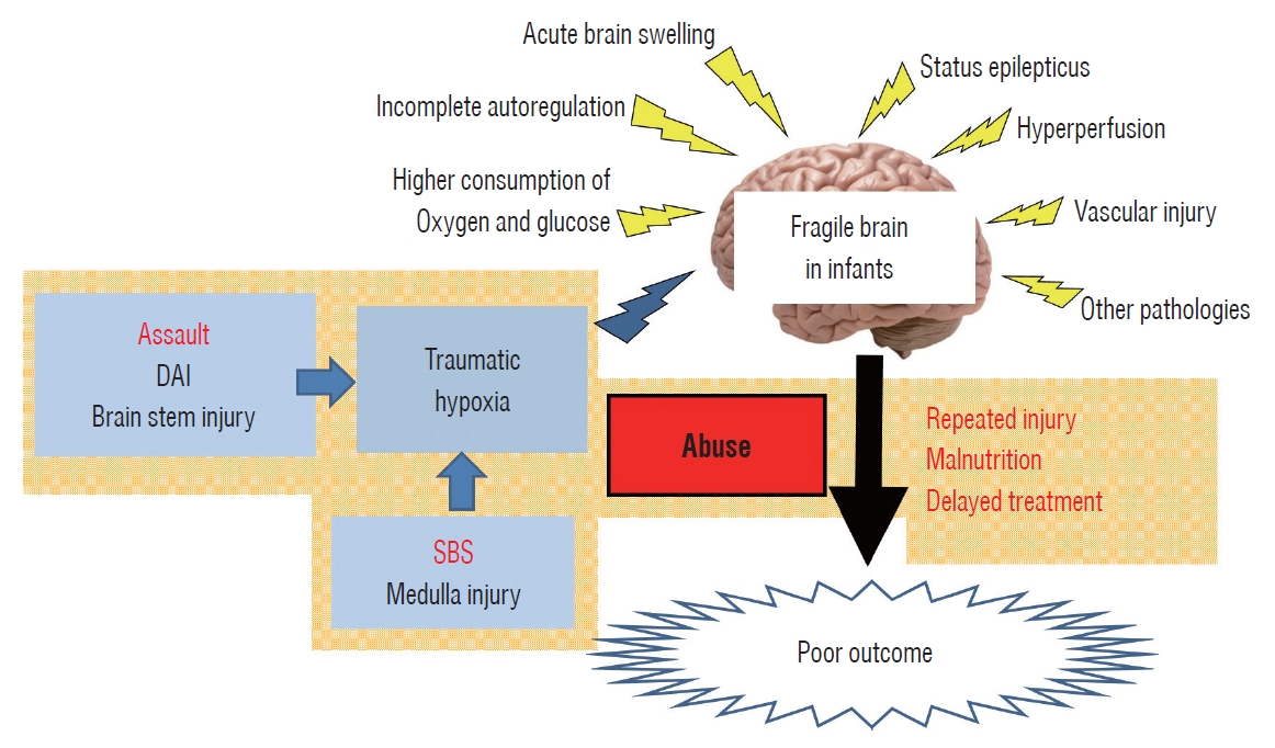
The main factors are summarized as follows. 1) Occurrence of eLDA discussed so far (this condition is not specific to AHT), 2) severe brain injury that causes CPA, specifically diffuse brain injury or direct damage to the medulla oblongata and upper cervical spinal cord, and 3) the social background peculiar to AHT. It should be considered that these factors are associated with in a complex manner (Fig. 14).
Occurrence of eLDA causes significant cerebral atrophy. Rapid and progressive cerebral atrophy can occur days to weeks after a head injury, which is extremely rare, not only in adults but also in children. Physiological features of an infant's brain include immature autoregulation, high oxygen consumption, and high glucose consumption. Their brains can be affected by traumatic primary brain injury and various secondary injuries caused by subdural hematomas, which can result in irreversible damage to the brain. Despite the expected growth and development of the brain, catastrophic damage occurs. In most cases, primary contusion injuries due to head trauma are absent or mild. Nevertheless, it should be recognized that AHT can cause brain atrophy and severe neurodevelopmental retardation.
Furthermore, when violent shaking is applied, in addition to respiratory disorders due to excessive passive movement of the cranio-cervical junction, extension vascular damage can affect the internal carotid artery and vertebro-basilar artery. This conception might be a new finding when considering the aggravation mechanism in AHT (Fig. 15).
Additionally, the specific problems of AHT cases are repeated physical assaults, malnutrition due to care neglect and delayed visits to the hospital, that is, medical neglect. Time is of the essence in treating head injuries. However, despite such emergencies, we have experienced many cases of delay in reporting to the hospital.
In cases of respiratory arrest or CPA at the time of transportation or visit, a poor outcome is inevitable regardless of the treatment.
Furthermore, all societal efforts to prevent AHT are extremely important [4,12,13].

CONCLUSION

The widespread LDA found in AHT head CT findings is not caused by a single pathology, but by the complex and overlapping additional effect of various harmful physiological factors to the brain of vulnerable infants. The pattern of eLDA that occurs days after injury is not appropriate to be considered a simple ischemic change. Pathological conditions such as status epilepticus and hyperperfusion injury are deeply involved. The pattern of eLDA that occurs early after a head injury is unlikely to occur except for severe disturbance of consciousness and hypoxic-ischemic changes due to CPA, and it is important to identify the cause. In-depth knowledge and abundant clinical experience of such pathological conditions are essential for appropriate AHT judgment.

Notes

Conflicts of interest

No potential conflict of interest relevant to this article was reported.

Informed consent

This type of study does not require informed consent.

Author contributions

Conceptualization : YSP; Data curation : YSP; Formal analysis : YSP; Methodology : YSP

Data sharing

None

Preprint

None

Fig. 1.
A : A typical computed tomography image of the so-called “Big Black Brain” showing hypodensity of the bilateral cerebral hemispheres. No lesions extend to the basal ganglia and thalamus. B : This case shows hypodensity of the entire cerebrum including the bilateral basal ganglia and thalamus on the initial computed tomography when transported to the hospital.
jkns-2021-0289f1.jpg
Fig. 2.
This case was transferred to the hospital with the chief complaint of cardiopulmonary arrest. Head computed tomography findings indicate that an extensive low-density area is widespread in the bilateral cerebrum, including the basal ganglia and thalamus, with no midline shift.
jkns-2021-0289f2.jpg
Fig. 3.
An extensive low-density area is observed in the right cerebral hemisphere with a severe midline shift. The amount of subdural hematoma is small.
jkns-2021-0289f3.jpg
Fig. 4.
A : Compute tomography on the day of abusive head trauma. B : Computed tomography performed 3 days after injury, a low-density area appeared locally appeared in the right frontal and parietal lobes (arrows).
jkns-2021-0289f4.jpg
Fig. 5.
A : Compute tomography on the day of abusive head trauma. B : Compute tomography performed 2 days after craniotomy, a low-density area locally appeared in the right frontal lobe and bilateral parieto-occipital lobes (arrows). These appearances are not due to a cerebral herniation.
jkns-2021-0289f5.jpg
Fig. 6.
A : Compute tomography (CT) on the day of head injury. B : Two days after craniotomy. CT shows an extensive low-density area in the right parietal lobe (arrow), fluid attenuated inversion recovery imaging shows diffuse high intensity in the right cerebral hemisphere, and arterial spin labeling imaging shows hyperperfusion at the same site.
jkns-2021-0289f6.jpg
Fig. 7.
Severe cerebral atrophy occurred 3 weeks after abusive head trauma (1-month-old boy).
jkns-2021-0289f7.jpg
Fig. 8.
A : A case of cardiopulmonary arrest (2-year-old girl) shows traumatic subarachnoid hemorrhage in the perimesencephalic cistern. B : A case of cardiopulmonary arrest (11-month-old girl) shows diffuse brain swelling immediately after head injury.
jkns-2021-0289f8.jpg
Fig. 9.
A case of cardiopulmonary arrest (4-month-old girl). A : Compute tomography shows acute subdural hematoma and typical hypoxic ischemic encephalopathy. B : Susceptibility weighted imaging image does not show traumatic primary damage to the brain parenchyma. C : Cervical cord magnetic resonance imaging also does not show traumatic lesions.
jkns-2021-0289f9.jpg
Fig. 10.
A : A case of cardiopulmonary arrest (6-month-old boy) shows retro clival hematoma, indirectly indicating medulla oblongata injury. B : A case of cardiopulmonary arrest (11-month-old girl) shows severe brain stem swelling immediately after injury.
jkns-2021-0289f10.jpg
Fig. 11.
A case of cardiopulmonary arrest (4-year-old boy) shows upper cervical cord stem swelling in the postmortem spine T2 weighted magnetic resonance imaging.
jkns-2021-0289f11.jpg
Fig. 12.
A cardiopulmonary arrest case (1-year-old girl) shows multiple trivial intracranial hematomata. However, there are no other serious primary brain parenchymal injuries. We conclude that the cause of cardiopulmonary arrest is asphyxia after vomiting.
jkns-2021-0289f12.jpg
Fig. 13.
Requirements for appropriate abusive head trauma judgment. SBS : shaken baby syndrome, ASDH : acute subdural hematoma, CT : computed tomography, MRI : magnetic resonance imaging, eLDA : extensive low-density area, BBB : big black brain, AHT : abusive head trauma.
jkns-2021-0289f13.jpg
Fig. 14.
Complex mechanism of poor outcome in infantile acute subdural hematoma, especially in abusive head trauma cases. DAI : diffuse axonal injury, SBS : shaken baby syndrome.
jkns-2021-0289f14.jpg
Fig. 15.
A : A 4-month-old girl with severe disturbance of consciousness. Computed tomography (CT) shows a low-density area in both the right cerebral hemisphere and the anterior area of the left frontal lobe, implicating an impaired blood flow in the right internal carotid artery. B : A 3-month-old boy with cardiopulmonary arrest. CT shows a low-density area in not only the brain stem but also in the thalamus and occipital lobes bilaterally and was considered to be impaired blood flow in the basilar artery.
jkns-2021-0289f15.jpg

References

1. Abu Hamdeh S, Marklund N, Lewén A, Howells T, Raininko R, Wikström J, et al : Intracranial pressure elevations in diffuse axonal injury: association with nonhemorrhagic MR lesions in central mesencephalic structures. J Neurosurg 131 : 604-611, 2018
crossref pmid
2. Adams JH, Doyle D, Ford I, Gennarelli TA, Graham DI, McLellan DR : Diffuse axonal injury in head injury: definition, diagnosis and grading. Histopathology 15 : 49-59, 1989
crossref pmid
3. Adamsbaum C, Grabar S, Mejean N, Rey-Salmon C : Abusive head trauma: judicial admissions highlight violent and repetitive shaking. Pediatrics 126 : 546-555, 2010
crossref pmid
4. Altman RL, Canter J, Patrick PA, Daley N, Butt NK, Brand DA : Parent education by maternity nurses and prevention of abusive head trauma. Pediatrics 128 : e1164-e1172, 2011
crossref pmid
5. Arndt DH, Lerner JT, Matsumoto JH, Madikians A, Yudovin S, Valino H, et al : Subclinical early posttraumatic seizures detected by continuous EEG monitoring in a consecutive pediatric cohort. Epilepsia 54 : 1780-1788, 2013
crossref pmid pmc
6. Brown RL, Brunn MA, Garcia VF : Cervical spine injuries in children: a review of 103 patients treated consecutively at a level 1 pediatric trauma center. J Pediatr Surg 36 : 1107-1114, 2001
crossref pmid
7. Choudhary AK, Servaes S, Slovis TL, Palusci VJ, Hedlund GL, Narang SK, et al : Consensus statement on abusive head trauma in infants and young children. Pediatr Radiol 48 : 1048-1065, 2018
crossref pmid
8. Christian CW, Block R, Committee on Child Abuse and Neglect; American Academy of Pediatrics : Abusive head trauma in infants and children. Pediatrics 123 : 1409-1411, 2009
crossref pmid
9. Colombari M, Troakes C, Turrina S, Tagliaro F, De Leo D, Al-Sarraj S : Spinal cord injury as an indicator of abuse in forensic assessment of abusive head trauma (AHT). Int J Legal Med 135 : 1481-1498, 2021
crossref pmid pmc
10. Cordobes F, Lobato RD, Rivas JJ, Portillo JM, Sarabia M, Munoz MJ : Post-traumatic diffuse brain swelling: isolated or associated with cerebral axonal injury. Clinical course and intracranial pressure in 18 children. Childs Nerv Syst 3 : 235-238, 1987
crossref pmid
11. Costine-Bartell BA, McGuone D, Price G, Crawford E, Keeley KL, Munoz-Pareja J, et al : Development of a model of hemispheric hypodensity ("big black brain"). J Neurotrauma 36 : 815-833, 2019
crossref pmid pmc
12. Dias MS, Rottmund CM, Cappos KM, Reed ME, Wang M, Stetter C, et al : Association of a postnatal parent education program for abusive head trauma with subsequent pediatric abusive head trauma hospitalization rates. JAMA Pediatr 171 : 223-229, 2017
crossref pmid pmc
13. Dias MS, Smith K, DeGuehery K, Mazur P, Li V, Shaffer ML : Preventing abusive head trauma among infants and young children: a hospital-based, parent education program. Pediatrics 115 : e470-e477, 2005
crossref pmid
14. Duhaime AC, Bilaniuk L, Zimmerman R : The “big black brain”: radiographic changes after severe inflicted head injury in infancy. J Neurotrauma 10(Suppl 1):S59, 1993

15. Duhaime AC, Christian C, Moss E, Seidl T : Long-term outcome in infants with the shaking-impact syndrome. Pediatr Neurosurg 24 : 292-298, 1996
crossref pmid
16. Duhaime AC, Christian CW : Abusive head trauma: evidence, obfuscation, and informed management. J Neurosurg Pediatr 24 : 481-488, 2019
crossref pmid
17. Duhaime AC, Christian CW, Rorke LB, Zimmerman RA : Nonaccidental head injury in infants--the “shaken-baby syndrome”. N Engl J Med 338 : 1822-1829, 1988
crossref
18. Duhaime AC, Durham S : Traumatic brain injury in infants: the phenomenon of subdural hemorrhage with hemispheric hypodensity (“big black brain”). Prog Brain Res 161 : 293-302, 2007
crossref pmid
19. Eder HG, Legat JA, Gruber W : Traumatic brain stem lesions in children. Childs Nerv Syst 16 : 21-24, 2000
crossref pmid
20. Figueira Rodrigues Vieira G, Guedes Correa JF : Early computed tomography for acute post-traumatic diffuse axonal injury: a systematic review. Neuroradiology 62 : 653-660, 2020
crossref pmid pmc
21. Foster KA, Recker MJ, Lee PS, Bell MJ, Tyler-Kabara EC : Factors associated with hemispheric hypodensity after subdural hematoma following abusive head trauma in children. J Neurotrauma 31 : 1625-1631, 2014
crossref pmid pmc
22. Gennarelli TA, Thibault LE, Adams JH, Graham DI, Thompson CJ, Marcincin RP : Diffuse axonal injury and traumatic coma in the primate. Ann Neurol 12 : 564-574, 1982
crossref pmid
23. Gerber P, Coffman K : Nonaccidental head trauma in infants. Childs Nerv Syst 23 : 499-507, 2007
crossref pmid
24. Graupman P, Winston KR : Nonaccidental head trauma as a cause of childhood death. J Neurosurg 104(4 Suppl):245-250, 2006
crossref pmid
25. Greeley CS : Abusive head trauma: a review of the evidence base. AJR Am J Roentgenol 204 : 967-973, 2015
crossref pmid
26. Greiner MV, Greiner HM, Caré MM, Owens D, Shapiro R, Holland K : Adding insult to injury: nonconvulsive seizures in abusive head trauma. J Child Neurol 30 : 1778-1784, 2015
crossref pmid pdf
27. Hasbani DM, Topjian AA, Friess SH, Kilbaugh TJ, Berg RA, Christian CW, et al : Nonconvulsive electrographic seizures are common in children with abusive head trauma*. Pediatr Crit Care Med 14 : 709-715, 2013
crossref pmid pmc
28. Karibe H, Kameyama M, Hayashi T, Narisawa A, Tominaga T : Acute subdural hematoma in infants with abusive head trauma: a literature review. Neurol Med Chir (Tokyo) 56 : 264-273, 2016
crossref pmid pmc
29. Khan NR, Fraser BD, Nguyen V, Moore K, Boop S, Vaughn BN, et al : Pediatric abusive head trauma and stroke. J Neurosurg Pediatr 20 : 183-190, 2017
crossref pmid
30. Kim HJ : The prognostic factors related to traumatic brain stem injury. J Korean Neurosurg Soc 51 : 24-30, 2012
crossref pmid pmc
31. Lee WJ, Lim YC, Yoon SH : Abusive head traumas in 4 infants. Korean J Neurotrauma 16 : 246-253, 2020
crossref pmid pmc
32. Mata-Mbemba D, Mugikura S, Nakagawa A, Murata T, Ishii K, Kushimoto S, et al : Traumatic midline subarachnoid hemorrhage on initial computed tomography as a marker of severe diffuse axonal injury. J Neurosurg 129 : 1317-1324, 2018
crossref pmid
33. Mata-Mbemba D, Mugikura S, Nakagawa A, Murata T, Kato Y, Tatewaki Y, et al : Intraventricular hemorrhage on initial computed tomography as marker of diffuse axonal injury after traumatic brain injury. J Neurotrauma 32 : 359-365, 2015
crossref pmid
34. Moen KG, Brezova V, Skandsen T, Håberg AK, Folvik M, Vik A : Traumatic axonal injury: the prognostic value of lesion load in corpus callosum, brain stem, and thalamus in different magnetic resonance imaging sequences. J Neurotrauma 31 : 1486-1496, 2014
crossref pmid
35. Narang SK, Estrada C, Greenberg S, Lindberg D : Acceptance of shaken baby syndrome and abusive head trauma as medical diagnoses. J Pediatr 177 : 273-278, 2016
crossref pmid
36. Narang SK, Fingarson A, Lukefahr J, COUNCIL ON CHILD ABUSE AND NEGLECT : Abusive head trauma in infants and children. Pediatrics 145 : e202002032020
crossref pmid
37. O'Neill BR, Handler MH, Tong S, Chapman KE : Incidence of seizures on continuous EEG monitoring following traumatic brain injury in children. J Neurosurg Pediatr 16 : 167-176, 2015
crossref pmid
38. Onuma T, Shimosegawa Y, Kameyama M, Arai H, Ishii K : Clinicopathological investigation of gyral high density on computerized tomography following severe head injury in children. J Neurosurg 82 : 995-1001, 1995
crossref pmid
39. Paek D, Kwon DI : A review on four different paths to respiratory arrest from brain injury in children; implications for child abuse. J Forensic Leg Med 71 : 101938, 2020
crossref pmid
40. Pang D : Spinal cord injury without radiographic abnormality in children, 2 decades later. Neurosurgery 55 : 1325-1342; discussion 1342-1343, 2004
crossref pmid
41. Pang D, Wilberger JE Jr : Spinal cord injury without radiographic abnormalities in children. J Neurosurg 57 : 114-129, 1982
crossref pmid
42. Piteau SJ, Ward MG, Barrowman NJ, Plint AC : Clinical and radiographic characteristics associated with abusive and nonabusive head trauma: a systematic review. Pediatrics 130 : 315-323, 2012
crossref pmid
43. Rongchao S, Shudong Y, Zhiyi Z : Pathological and immunohistochemical study of lethal primary brain stem injuries. Diagn Pathol 7 : 54, 2012
crossref pmid pmc
44. Visocchi M, Chiaretti A, Genovese O, Di Rocco F : Haemodynamic patterns in children with posttraumatic diffuse brain swelling. A preliminary study in 6 cases with neuroradiological features consistent with diffuse axonal injury. Acta Neurochir (Wien) 149 : 347-356, 2007
crossref pmid
45. Wong AM, Yeh CH, Liu HL, Wu TW, Lin KL, Wang HS, et al : Arterial spin-labeling perfusion imaging of children with subdural hemorrhage: perfusion abnormalities in abusive head trauma. J Neuroradiol 44 : 281-287, 2017
crossref pmid
46. Yokobori S, Nakae R, Yokota H, Spurlock MS, Mondello S, Gajavelli S, et al : Subdural hematoma decompression model: a model of traumatic brain injury with ischemic-reperfusional pathophysiology: a review of the literature. Behav Brain Res 15 : 23-28, 2018
crossref
47. Zimmerman RA, Bilaniuk LT, Farina L : Non-accidental brain trauma in infants: diffusion imaging, contributions to understanding the injury process. J Neuroradiol 34 : 109-114, 2007
crossref pmid
Editorial Office
1F, 18, Heolleung-ro 569-gil, Gangnam-gu, Seoul, Republic of Korea
TEL: +82-2-525-7552   FAX: +82-2-525-7554   E-mail: office@jkns.or.kr
About |  Browse Articles |  Current Issue |  For Authors and Reviewers
Copyright © Korean Neurosurgical Society.                 Developed in M2PI
Close layer