|
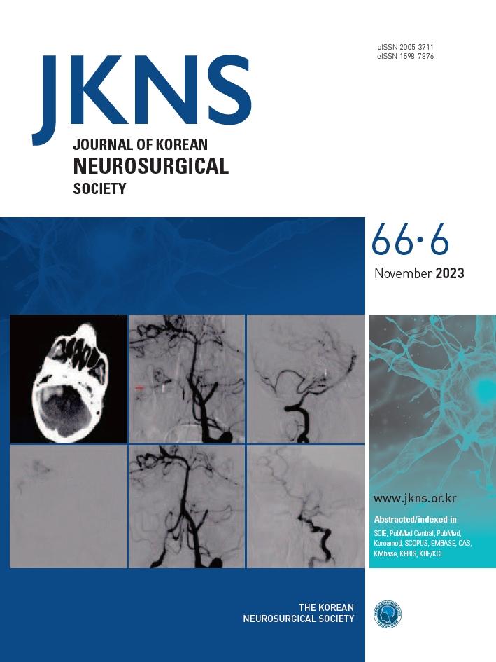
Intracranial dural arteriovenous fistula (dAVF) resulting in hemorrhage in the posterior fossa. Upper left: right cerebellar hematoma on noncontrast cranial computed tomography. Upper center and right : left vertebral artery digital subtraction angiogram shows a dAVF (white arrow) fed from the right pial supply from the anterior inferior cerebellar artery (AICA, black arrow), draining into the superior petrosal sinus (red arrow). Lower left: embolization with Onyx after microcatheterization via AICA. Lower center and right : complete closure of the fistula in the post-operative first year of follow-up.
|