|
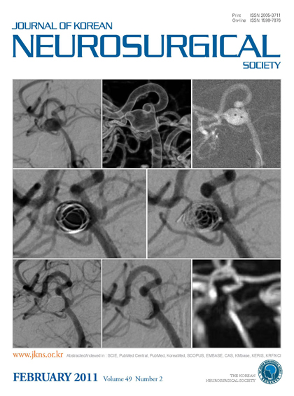
Case 9; a 63-year-old women with SAH. A : The left vertebral artery angiogram shows the aneurysm before treatment. B : 3D reconstruction image shows wide-necked right superior cerebellar artery aneurysm (neck×height×width×length : 5.0×7.1×5.3×6.2 mm). C : Two microcatheters are positioned within the aneurysm. One has a 45° angled distal tip and the other has a 90° angled distal tip. D : First coil (360° Guglielmi detachable coil 7×15 mm) being deployed. E : Second coil (Microplex coil Complex 5×15 mm) being deployed via another microcatheter. F : Immediately after embolization, angiogram shows compact occlusion of the aneurysmal sac and patent right SCA. There is a small remnant neck around the right SCA. G : Two months after embolization, an angiogram shows no interval change and stable coils. H : Six months after embolization, magnetic resonance angiogram shows no interval change and stable coils.
|